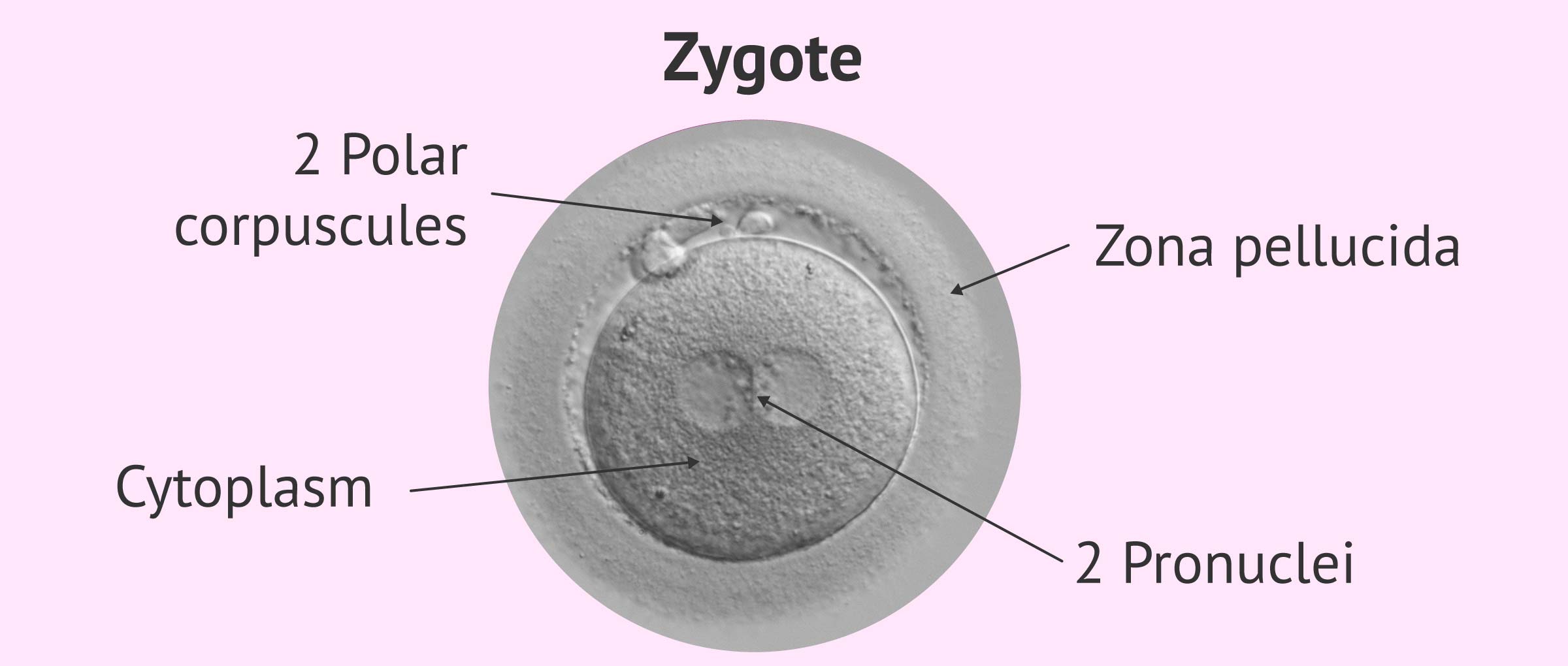
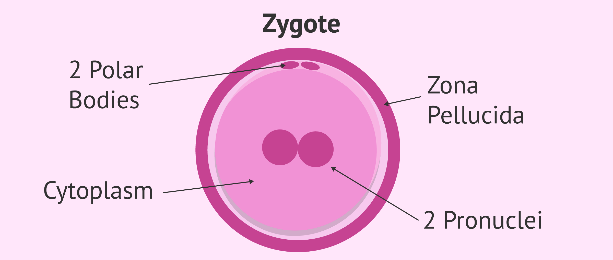
Zygote Drawing
Zygote Drawing - Zygote formation diagram / formation and development of an embryo from the zygote class 8 today i will show you zygote formation diagram. Web get your medical education, 3d anatomical and medical model questions answered here on our zygote faq section. All images photos vectors illustrations 3d objects. Overview of fertilization and early human development. Embryo development 19 century medical illustration. To ensure that each zygote has the correct number of chromosomes, only one sperm can fuse with one egg. The zygote stage development occurs in the first week of fertilization. Sperm and egg cells, known as gametes, fuse during fertilization to create a zygote. Web your first form as a zygote split to make two cells. Photograph of the original illustration from a system of human anatomy by erasmus wilson published in 1859. Why has zygote been the industry standard for so many years? The cellular mechanisms present in the gametes also function in the zygote, but the newly fused dna produces a different effect in the new cell. The zygote is formed when the male gamete (sperm) and female gamete (egg) fuse. Zygote formation diagram / formation and development of an embryo. Web browse 40+ human zygote drawing stock photos and images available, or start a new search to explore more stock photos and images. There are two ways cell division can happen in humans and. Web fertilization is the process in which haploid gametes fuse to form a diploid cell called a zygote. Want to join the conversation? A zygote is. Web choose from 41 human zygote drawing stock illustrations from istock. Gametes have half the chromosomes (haploid) of a typical body cell, while zygotes have the full set (diploid). 2.6k views 1 year ago class 8 science diagrams. Sperm and egg cells, known as gametes, fuse during fertilization to create a zygote. Embryo development 19 century medical illustration. Zygote formation diagram / formation and development of an embryo from the zygote class 8 today i will show you zygote formation diagram. Web browse 40+ human zygote drawing stock photos and images available, or start a new search to explore more stock photos and images. Photograph of the original illustration from a system of human anatomy by erasmus wilson published in 1859. A zygote is the cell formed when two gametes fuse during fertilization. There are two ways cell division can happen in humans and. 2.6k views 1 year ago class 8 science diagrams. The cellular mechanisms present in the gametes also function in the zygote, but the newly fused dna produces a different effect in the new cell. The zygote stage development occurs in the first week of fertilization. The dna material from the two cells is combined in the resulting zygote. Web your first form as a zygote split to make two cells. Overview of fertilization and early human development. Complete male and female collections or individual anatomical anatomy. Learn how a zygote, the single cell produced by fertilization, divides by mitosis to produce all the tissues of the human body (including germ cells, which can undergo meiosis to make sperm and eggs). Embryo development 19 century medical illustration. See zygote stock video clips. Want to join the conversation?
Zygote 3d Anatomy

Structure of Zygote

Zygote, illustration Stock Image C039/2357 Science Photo Library
Want To Join The Conversation?
Web Get Your Medical Education, 3D Anatomical And Medical Model Questions Answered Here On Our Zygote Faq Section.
Why Has Zygote Been The Industry Standard For So Many Years?
Web 3D Human Anatomy Models For Illustrators And Animators.
Related Post: