Synovial Joint Drawing
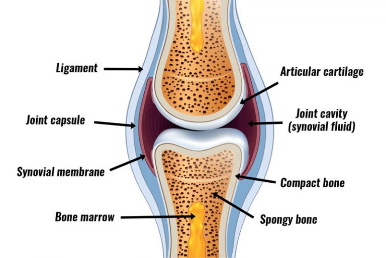
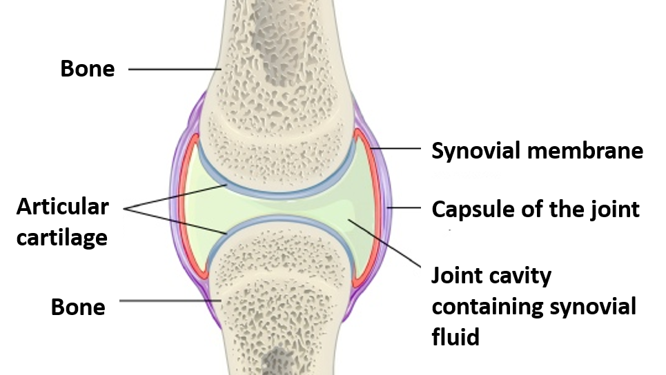
Synovial Joint Drawing - Synovial joints are the most common type of. List the six types of synovial joints and give an example of each. The bones of the joint articulate with each other within the joint cavity. Web 1 key structures of a synovial joint. The articulating surfaces of the bones are covered by a thin layer of articular cartilage. Describe the structural features of a synovial joint. There are 6 types of synovial joints. The articulating surfaces of the bones are covered by a thin layer of articular cartilage. Including what synovial fluid is, where the synovial membrane is, what joint capsules are, the ro. They have varying shapes, but the important thing about them is the movement they. Discuss the structure of specific body joints and the movements allowed by each. They have varying shapes, but the important thing about them is the movement they. List the six types of synovial joints and give an example of each. Synovial joints allow for smooth movements between the adjacent bones. This video shows how to draw the. This video shows how to draw the. A synovial membrane (or synovium) is the soft tissue found between the articular capsule (joint capsule) and the joint cavity of synovial joints. Synovial joints allow for smooth movements between the adjacent bones. They have varying shapes, but the important thing about them is the movement they. A key structural characteristic for a. From ‘human biology’ by d. Web a synovial joint is a connection between two bones consisting of a cartilage lined cavity filled with fluid, which is known as a diarthrosis joint. Describe the structures that support and stabilize each joint. Web describe the structural features of a synovial joint. Including what synovial fluid is, where the synovial membrane is, what. List the six types of synovial joints and give an example of each. Web a revision lesson on how to draw and label synovial joints. The articulating surfaces of the bones are covered by a thin layer of articular cartilage. The hinge joint is one of six types of synovial joints along with the plane, ellipsoid, ball and socket, pivot and saddle joints. Cavitas articularis (junctura synovialis) capsula. 4m views 9 years ago anatomy of the human body for artists | proko. Synovial joints achieve movement at the point of contact of the articulating bones. They have varying shapes, but the important thing about them is the movement they. The femur, tibia and patella. The joint is surrounded by an articular capsule that defines a joint cavity filled with synovial fluid. The tibiofemoral joint and patellofemoral joint. Synovial joints allow for smooth movements between the adjacent bones. This joint unites long bones and permits free bone movement and greater mobility. Web synovial joints are characterized by the presence of a joint cavity. Multiaxial joint , the articular surfaces are essentially flat, and they allow only short nonaxial gliding movements. There are six types of synovial joints.
Human synovial joint Download Scientific Diagram

Synovial Joint Structure

Synovial joints Anatomy QA
The Articulating Surfaces Of The Bones Are Covered By A Thin Layer Of Articular Cartilage.
Including What Synovial Fluid Is, Where The Synovial Membrane Is, What Joint Capsules Are, The Ro.
From ‘Human Biology’ By D.
Web A Synovial Joint, Also Known As Diarthrosis, Joins Bones Or Cartilage With A Fibrous Joint Capsule That Is Continuous With The Periosteum Of The Joined Bones, Constitutes The Outer Boundary Of A Synovial Cavity, And Surrounds The Bones' Articulating Surfaces.
Related Post: