Small Intestine Drawing
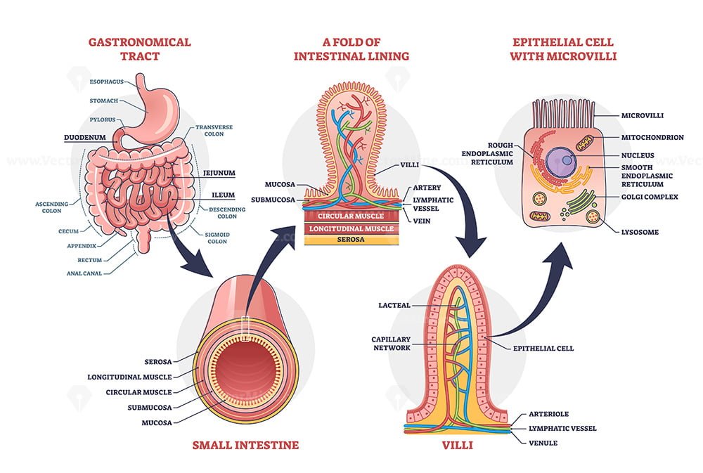
Small Intestine Drawing - Learn and reinforce your understanding of small intestine histology. Anatomy of the human digestive system with a description of the. The duodenum, where most digestion occurs, the jejunum, where nutrients are absorbed, and the ileum, where important vitamins are absorbed. Describe the histology of the small intestine. Old engraved illustration of human digestive system. Small intestine histology videos, flashcards, high yield notes, & practice questions. Together with the esophagus, large intestine, and the stomach, it forms the gastrointestinal tract. The small intestine or small bowel is an organ in the gastrointestinal tract where most of the absorption of nutrients from food takes place. The main functions of the small intestine are to complete digestion of food and to absorb nutrients. Also shown are the stomach, appendix, large intestine, and rectum. The intestines are responsible for breaking food down, absorbing its nutrients and solidifying the waste. Image information and view/download options. Anatomy of the human digestive system with a description of the. Drawing of the small intestine showing the duodenum, jejunum, and ileum. Explore the small intestine's role in digestion and nutrient absorption. The small intestine or small bowel is an organ in the gastrointestinal tract where most of the absorption of nutrients from food takes place. Learn and reinforce your understanding of small intestine histology. Web browse 138 small intestine drawing photos and images available, or start a new search to explore more photos and images. Discover the role of villi and. It lies between the stomach and large intestine, and receives bile and pancreatic juice through the pancreatic duct to aid in digestion. The small intestine consists of three parts: Learn about the three parts: Histology of the small intestine. Web how to draw small intestine. The outermost serosa, muscularis, submucosa, and innermost mucosa. New 3d rotate and zoom. Web see small intestine drawing stock video clips. The duodenum, where most digestion occurs, the jejunum, where nutrients are absorbed, and the ileum, where important vitamins are absorbed. Web the small intestine: An illustration of the small intestine with the duodenum, jejunum, and ileum labeled. Web the small intestine (or small bowel) is a convoluted tube that begins at the pylorus of the stomach and ends approximately twenty feet (six meters) further down at the ileocecal valve. Anatomy of the human digestive system with a description of the. The intestines are responsible for breaking food down, absorbing its nutrients and solidifying the waste. It extends from the stomach ( pylorus) to the large intestine ( cecum) and consists of three parts: Drawing of the small intestine showing the duodenum, jejunum, and ileum. Web the small intestine is a organ located in the gastrointestinal tract, which assists in the digestion and absorption of ingested food. Labelled diagram of small intestine. Discover the role of villi and microvilli in increasing surface area for digestion. Anatomically, the small bowel can be divided into three parts; Small intestine histology videos, flashcards, high yield notes, & practice questions.
Small intestine with scientific gastrointestinal structure outline

Three parts of small intestine diagram

Human Small Intestine. Colored Sketch Stock Vector Illustration of
Histology Of The Small Intestine.
The Duodenum, Jejunum And Ileum.
Web The Small Bowel Connects To The Large Bowel, Also Called The Large Intestine Or Colon.
Learn About The Three Parts:
Related Post: