Sarcomere Drawing
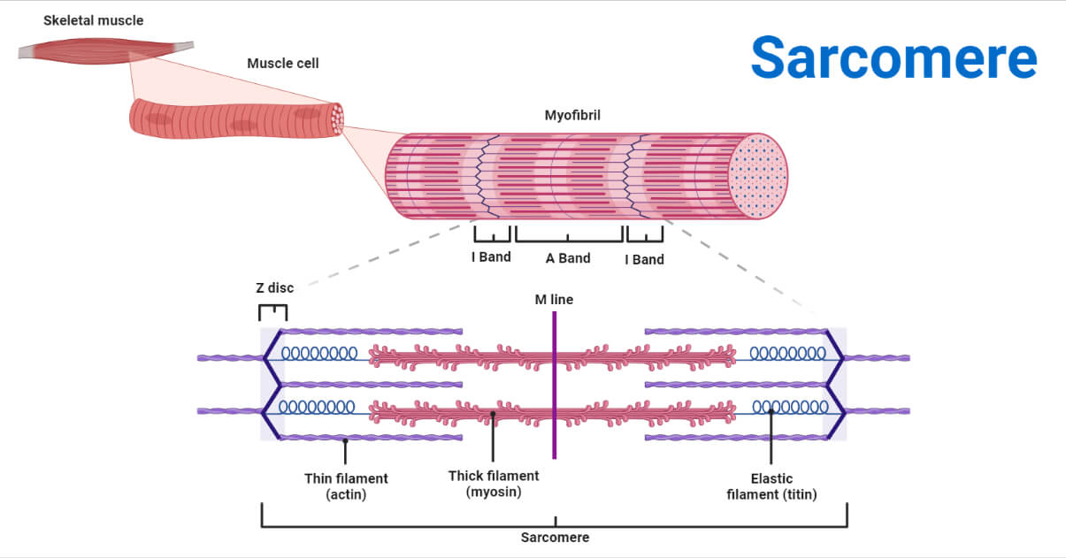
Sarcomere Drawing - Web a sarcomere (greek σάρξ sarx flesh, μέρος meros part) is the smallest functional unit of striated muscle tissue. List the major sarcomeric proteins involved with contraction; Actin myofilaments, which are shown in red, and myosin myofilaments, which are shown in blue. A sarcomere is the basic contractile unit of a myocyte (muscle fibre). Web the sarcomere is a main contractile unit of muscle fiber in the skeletal muscle. Herein lies the sarcomere’s main purpose. Control of the skeletal muscle contraction. By identifying these structures, one can gain a better understanding of how sarcomeres function and how muscle contractions occur. Web a labeled sarcomere diagram is a visual representation of the structural organization of a sarcomere, which is the fundamental unit of a muscle fiber. The sarcomere itself is bundled within the myofibril that runs the entire length of the muscle fiber and attaches to the sarcolemma at its end. The function of sarcolemma is to connect the basement membrane which wrapped all connective tissues. Sarcomeres play a crucial role in muscle contraction and their detailed study is essential in. The sarcomere fundamentally consists of two main myofilaments: Web a labeled sarcomere diagram is a visual representation of the structural organization of a sarcomere, which is the fundamental unit of. (b) a conceptual diagram representing the. Web the sarcomere is the functional unit of the muscle fiber. It also allows us to understand the visible bands seen in the images of muscle tissue in micrographs. Web (a) the basic organization of a sarcomere subregion, showing the centralized location of myosin (a band). Actin and the z discs are shown in. These filaments interact by sliding past each other in. Web the sarcomere is the functional unit of the muscle fiber. A sarcomere is the basic contractile unit of a myocyte (muscle fibre). Web the smallest unit of contraction is the sarcomere, where actin and myosin filaments interact to cause muscle contraction. Sarcomere muscular biology scheme vector illustration. As myofibrils contract, the entire muscle cell contracts. Thick filaments called myosin and thin filaments called actin. As we can see in this image of a sarcomere, it is made up of two main filaments: Web (a) the basic organization of a sarcomere subregion, showing the centralized location of myosin (a band). Web a sarcomere is a microscopic segment repeating in a myofibril. The sarcomere fundamentally consists of two main myofilaments: The left side (peach color) of the sarcomere represents a half sarcomere found in vertebrate skeletal myofibrils. Web a labeled sarcomere diagram is a visual representation of the structural organization of a sarcomere, which is the fundamental unit of a muscle fiber. Sarcomeres play a crucial role in muscle contraction and their detailed study is essential in. It also allows us to understand the visible bands seen in the images of muscle tissue in micrographs. Skeletal muscles are composed of tubular muscle cells (called muscle fibers or myofibers) which are formed during embryonic myogenesis. The myosin filaments are the thick filaments and should be represented as being thicker than the actin filaments. The function of sarcolemma is to connect the basement membrane which wrapped all connective tissues. Web the sarcomere is a main contractile unit of muscle fiber in the skeletal muscle. Control of the skeletal muscle contraction. Note that the nebulin molecules are part of and extend the entrie length of the thin filaments.
Diagram Diagram Quizlet
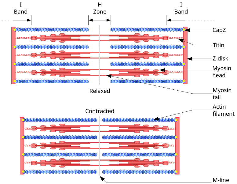
Diagram Of A
Definition, Structure, Diagram, and Functions
Actin And The Z Discs Are Shown In Red.
Web When Drawing A Diagram Of A Sarcomere It Is Important To Remember The Following Conventions:
Hence, Its Main Function Is The Regulation Of Muscle Contraction.
Web A Sarcomere (Greek Σάρξ Sarx Flesh, Μέρος Meros Part) Is The Smallest Functional Unit Of Striated Muscle Tissue.
Related Post: