Reticular Connective Tissue Drawing
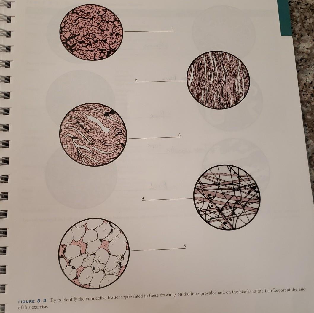
Reticular Connective Tissue Drawing - *font labels changed to red for easier visualization because the slide was stained dark. This scaffolding supports other cell types including white blood cells, mast cells, and macrophages. Web reticular connective tissue is a type of loose connective tissue that is abundant with reticular fibers. A slide of reticular connective tissue from a human spleen. Reticular cells are specialized fibroblasts that synthesize and hold the fibers. May anchor to collagenous septa, which divide organs into lobes. Web reticular connective tissues are arranged along with different cells in various organs like bone marrow, lymph nodes, spleen, liver, kidneys, and even under the skin. Reticular fibers are abundant in lymphoid organs (lymph nodes, spleen), bone marrow and liver. Reticular connective tissue forms a scaffolding for other cells in several organs, such as lymph nodes and bone marrow. Reticular fibers are composed of thin and delicately woven strands of type iii collagen. Reticular fibers are abundant in lymphoid organs (lymph nodes, spleen), bone marrow and liver. Found in lymph nodes, spleen, and bone marrow. Reticular connective tissue forms a scaffolding for other cells in several organs, such as lymph nodes and bone marrow. Web reticular connective tissue is a type of loose connective tissue that is abundant with reticular fibers. These soft. The cells that make the reticular fibers are fibroblasts called reticular cells. Forms stroma of liver, spleen, bone marrow, and lymph nodes. Web reticular connective tissues are arranged along with different cells in various organs like bone marrow, lymph nodes, spleen, liver, kidneys, and even under the skin. Fine reticular fibers stain faintly; These fibers are actually type iii collagen. The cells that make the reticular fibers are fibroblasts called reticular cells. Web reticular tissue is a special type of connective tissue that predominates in various locations that have a high cellular content. Web reticular connective tissue is a type of connective tissue with a network of reticular fibers, made of type iii collagen (reticulum = net or network). Reticular. Reticular connective tissue is named for the reticular fibers which are the main structural part of the tissue. *font labels changed to red for easier visualization because the slide was stained dark. Web reticular connective tissue forms an internal scaffolding for certain organs, such as lymph nodes, bone marrow, and the spleen. Identify the different cells and fiber types found in connective tissue. Appearance and features of the reticular connective tissue. These fibers are actually type iii collagen fibrils. Reticular cells are specialized fibroblasts that synthesize and hold the fibers. Found in lymph nodes, spleen, and bone marrow. Web reticular connective tissue is a type of connective tissue with a network of reticular fibers, made of type iii collagen (reticulum = net or network). A slide of reticular connective tissue from a human spleen. They are not visible with hematoxylin & eosin (h&e), but are specifically stained by silver. Reticular tissue, a type of loose connective tissue in which reticular fibers are the most prominent fibrous component, forms the supporting framework of the lymphoid organs (lymph nodes, spleen, tonsils), bone marrow and liver. If there is little space between protein fibers, the tissue is likely one of the dense connective tissues. Web o correlate the histological compositions and organizations of ct proper, reticular ct, and adipose ct and their locations and functions. Reticular fibers are not unique to reticular connective tissue, but only in this tissue type are they dominant. Dense regular connective tissue, including tendon and elastic (yellow) ligament.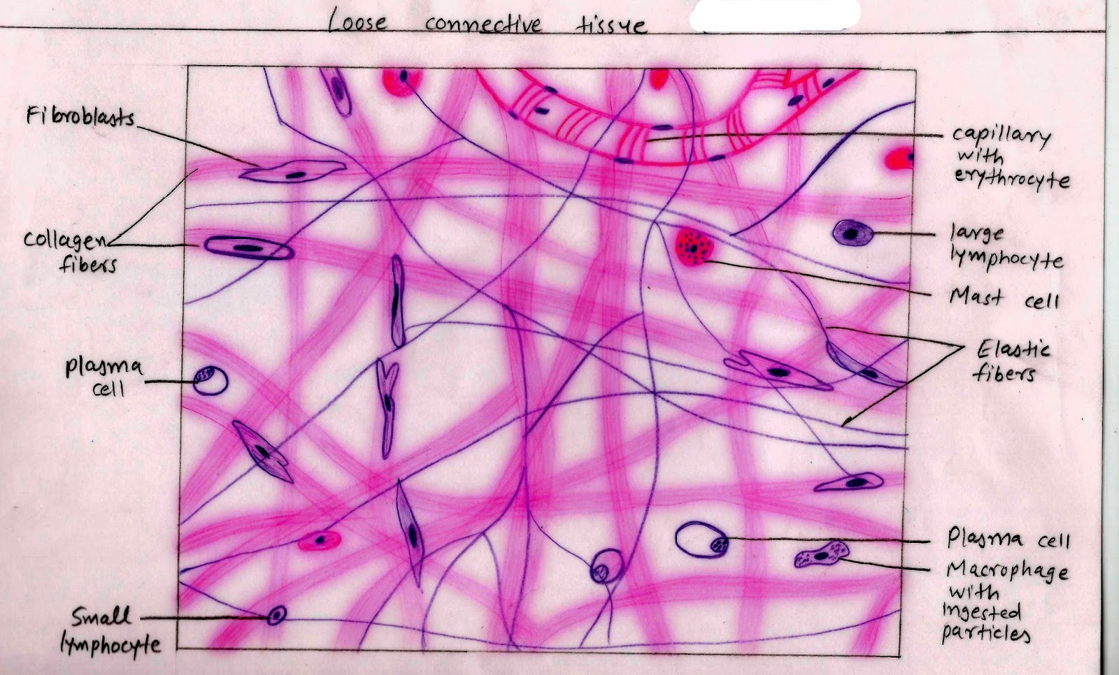
Histology Image Connective tissue
Reticular Connective Tissue Drawing Master the Art of Illustrating

Reticular Connective Tissue Structure
Function Of Reticular Connective Tissue.
Watch The Video Tutorial Now.
Reticular Fibers Are Abundant In Lymphoid Organs (Lymph Nodes, Spleen), Bone Marrow And Liver.
This Special Connective Tissue Forms The Stroma For Hemopoietic Tissues And Lymphoid Structures And Organs, Except The Thymus.
Related Post: