Prokaryotic Cell Drawing
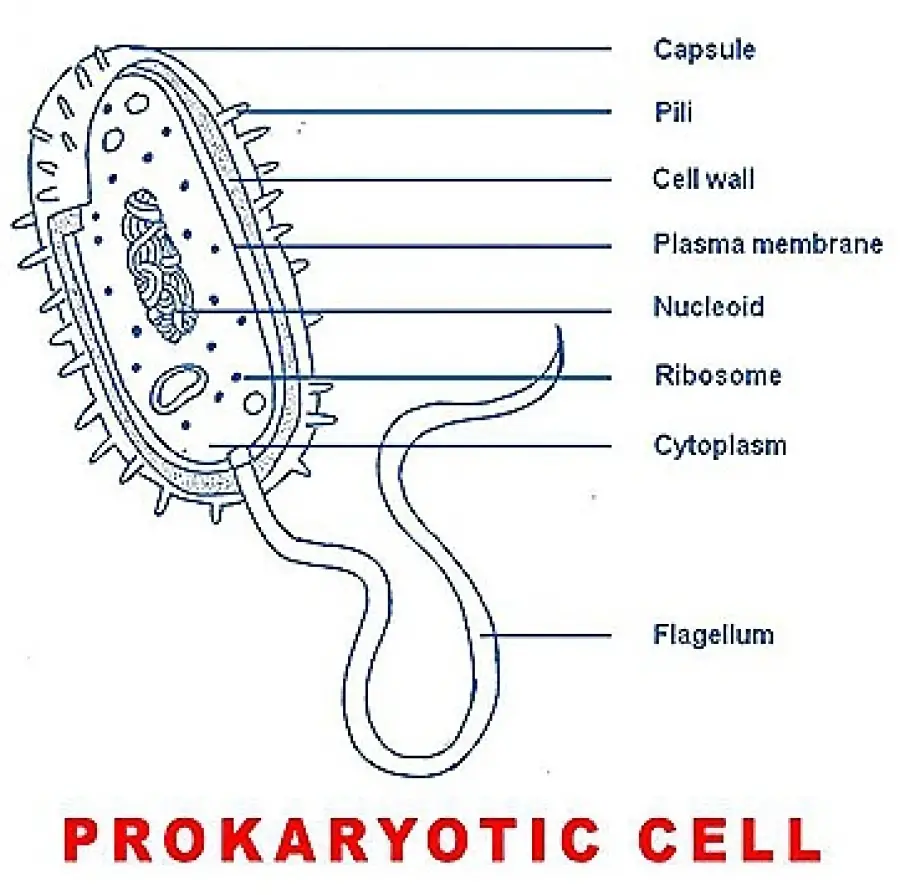
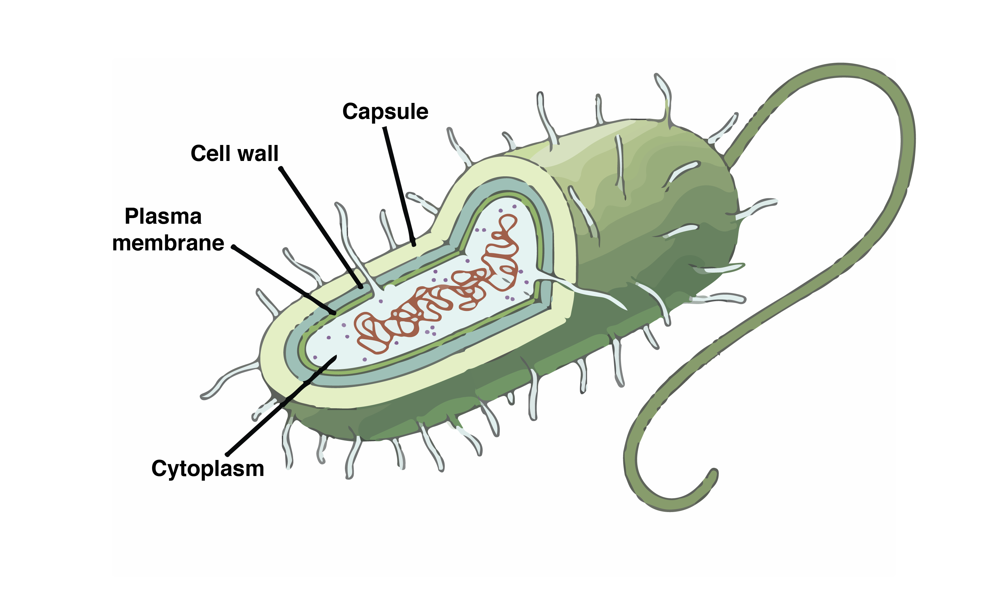
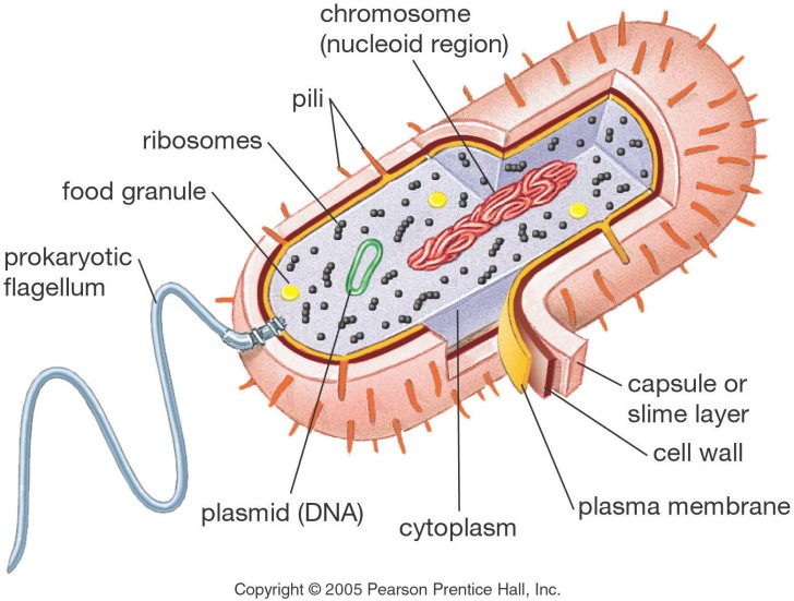
Prokaryotic Cell Drawing - Updated on october 30, 2019. Archaeal membranes have replaced the fatty acids of bacterial membranes with isoprene; Color a typical prokaryote cell. Definition, characteristics, diagram & structure. As organized in the three domain system, prokaryotes include bacteria and archaeans. The structure called a mesosome was once thought to be an organelle. The following image is a diagram of a prokaryotic cell; Compare and contrast prokaryotic cells and eukaryotic cells. Web cell wall, capsule, flagellum, phospholipids, murein / glycoprotein / peptidoglycans, cell surface membrane. Web general biology 1e (openstax) unit ii: This image shows a braarudosphaera bigelowii cell. The diagram of prokaryotic cell show that it contains genetic material in a nucleoid region, have a cell wall, and may possess flagella. These neat, well labelled and colorful diagrams will make your answers look more. The anatomy of a bacterial cell prokaryotic cell structure. Web typical prokaryotic cells range from 0.1 to. How to draw prokaryotic cell step by. Prokaryotic cells do not have a true nucleus that contains their genetic material as eukaryotic cells do. The main parts of a prokaryotic cell are shown in this diagram. Web general biology 1e (openstax) unit ii: Web typical prokaryotic cells range from 0.1 to 5.0 micrometers (μm) in diameter and are significantly smaller. The anatomy of a bacterial cell prokaryotic cell structure. Web the prokaryotic cell diagram given below represents a bacterial cell. Cell wall, capsule, flagellum, phospholipids, murein / glycoprotein / peptidoglycans, cell surface membrane. Schematic diagram of a prokaryotic cell. Web typical prokaryotic cells range from 0.1 to 5.0 micrometers (μm) in diameter and are significantly smaller than eukaryotic cells, which. And as you can imagine, shape may have something to do with mobility. In the following sections, we’ll walk through the structure of a prokaryotic cell, starting on the outside and moving towards the inside of the cell. The diagram of prokaryotic cell show that it contains genetic material in a nucleoid region, have a cell wall, and may possess flagella. Web i am demonstrating the colorful diagram of prokaryotic cells step by step which you can draw very easily. Web many prokaryotic cells have sphere, rod, or spiral shapes (as shown below). Structure and function of the plasma membrane and cytoplasm of cells. Web general biology 1e (openstax) unit ii: The cell is the basic unit of life and a cell can be either a prokaryotic cell or eukaryotic cell. Archaeal membranes have replaced the fatty acids of bacterial membranes with isoprene; Most prokaryotic cells are much smaller than eukaryotic cells. To know this, first, we should know what a cell is. Web typical prokaryotic cells range from 0.1 to 5.0 micrometers (μm) in diameter and are significantly smaller than eukaryotic cells, which usually have diameters ranging from 10 to 100 μm. These cells are structurally simpler and smaller than their eukaryotic counterparts, the cells that make up fungi, plants, and animals. Definition, characteristics, diagram & structure. Web kateryna kon/science photo library / getty images. Web prokaryotic cell diagram to help you remember prokaryotes parts and pieces.
Simple Prokaryotic Cell Diagram

Learning Through Art Structures of a Prokaryotic Cell

prokaryotic cell diagram Biological Science Picture Directory
Updated On October 30, 2019.
The Plasma Membrane—The Outer Boundary Of The Cell—Is The Bag, And The Cytoplasm Is The Goo.
In This Case, A Bacterium.
A Prokaryote Is A Unicellular Organism That Lacks A.
Related Post: