Neuron Drawing Labeled
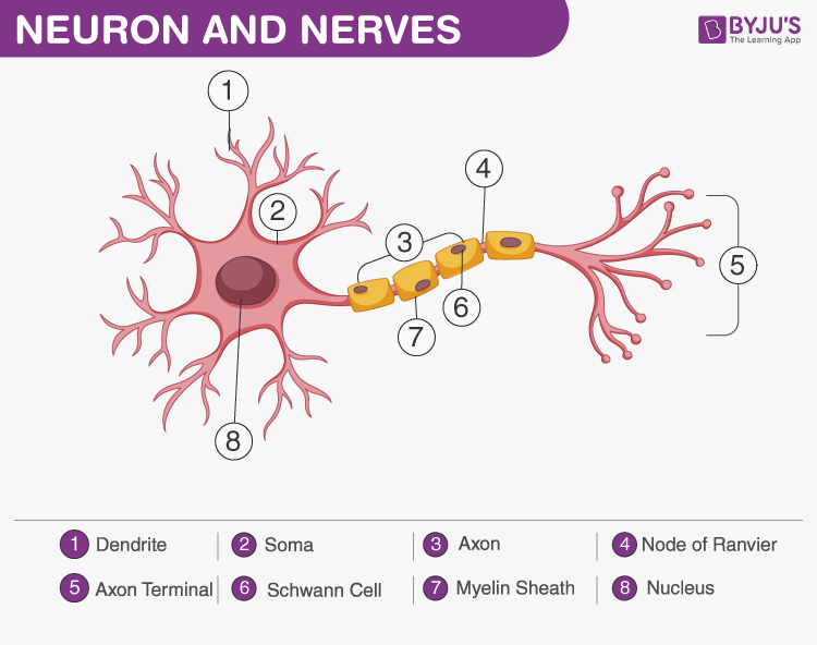
Neuron Drawing Labeled - Web what is a neuron? In the figure, labeled ‘2’ is the short filaments from the cell body that carries impulses from dendrites to the cell body which is the Neurons pass messages to each other using a special type of. Learn vocabulary, terms, and more with flashcards, games, and other study tools. Learn about the axon hillock, axon terminals, and the myelin sheath. The message then moves through the axon to the other end of the neuron, then to the tips of the axon and then into the space between neurons. Somatic motor neuron with cell body, axon, axon, myelin sheath, nodes of ranvier, axon terminal, dendrites, synaptic end of the bulbs, and other associated. Web the parts of the neuron have been labeled. Web a neuron is a nerve cell that processes and transmits information through electrical and chemical signals in the nervous system. The neuron doctrine is the now fundamental idea that neurons are the basic structural and functional units of the nervous system. At a synapse, one neuron sends a message to a target neuron—another cell. From there the message can move to the next neuron. Web start studying label parts of a neuron. The neuron doctrine is the now fundamental idea that neurons are the basic structural and functional units of the nervous system. A detailed diagram of a neuron, showing the. These synapses communicate using chemical messengers. The main structure of a neuron includes the following parts: If you need some help, visit the web article listed below. Neurons (or nerve cells) are specialized cells that transmit and receive electrical signals in the body. The message then moves through the axon to the other end of the neuron, then to the. Web start studying label parts of a neuron. Neurons pass messages to each other using a special type of. Web figure 12.8 parts of a neuron the major parts of the neuron are labeled on a multipolar neuron from the cns. Web a major challenge in explainable ai is in correctly interpreting activations of hidden neurons: Neurons are the structural. Neurons (or nerve cells) are specialized cells that transmit and receive electrical signals in the body. Web the parts of the neuron have been labeled. In these synapses, ions flow directly between cells. If you need some help, visit the web article listed below. Where the axon emerges from the cell body, there is a special region referred to as the axon hillock. Web from memory, draw a neuron and identify the following structures: The neuron doctrine is the now fundamental idea that neurons are the basic structural and functional units of the nervous system. Figure \(\pageindex{2}\) shows the structure of a typical neuron. They are found in the brain, spinal cord and the peripheral nerves. Neurons consist of a cell body, dendrites (which receive signals), and an axon (which sends signals). A detailed diagram of a neuron, showing the synapse, cell. Learn about the axon hillock, axon terminals, and the myelin sheath. Web find out what are the 3 different parts of a neuron and which part does what. It will also cover briefly the histological layers of the central and peripheral nervous systems. Explore the structure of neurons, their types, and functions. At a synapse, one neuron sends a message to a target neuron—another cell.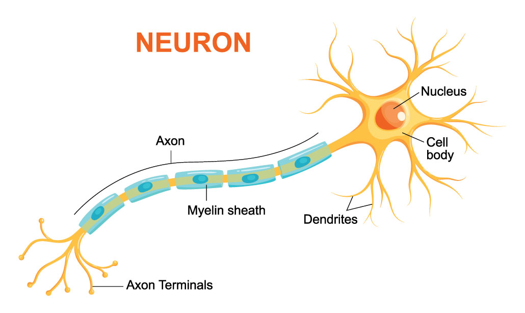
Explainer What is a neuron? Science News for Students
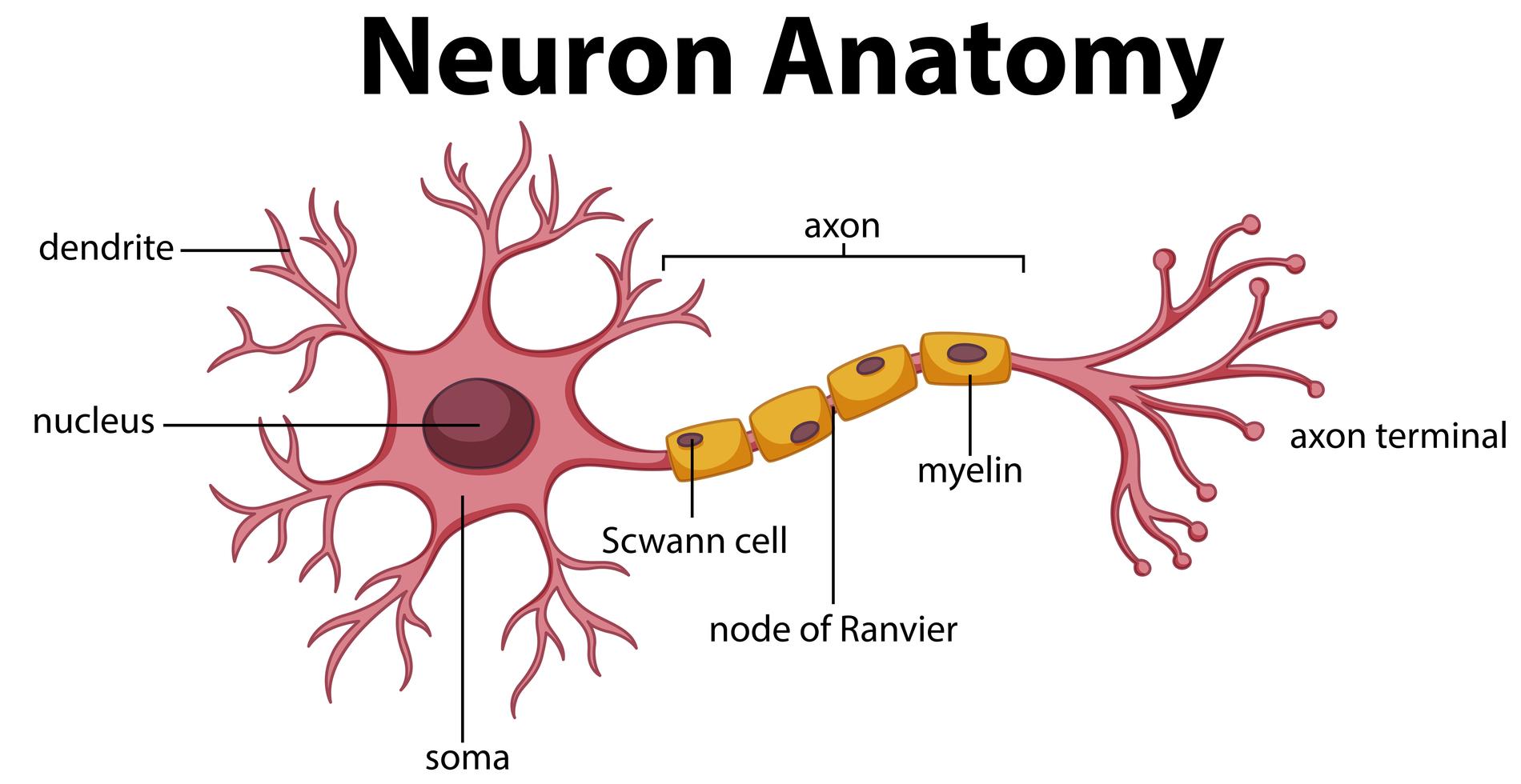
Diagram of Neuron Anatomy 358962 Vector Art at Vecteezy
What does a neuron look like? Biology Questions
These Synapses Communicate Using Chemical Messengers.
In The Figure, Labeled ‘2’ Is The Short Filaments From The Cell Body That Carries Impulses From Dendrites To The Cell Body Which Is The
Neurons Communicate With One Another At Junctions Called Synapses.
This Article Will Explain The Histology Of Neurons, Providing You With Information About Their Structure, Types, And Clinical Relevance.
Related Post: