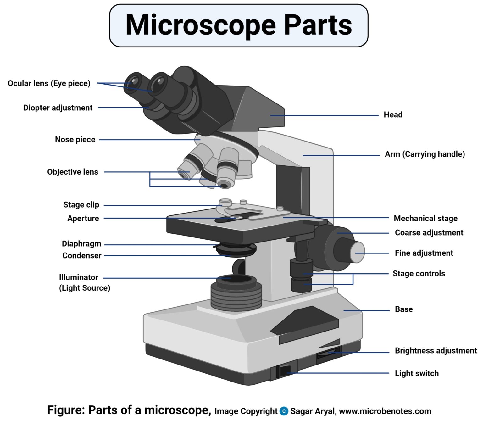
Labelled Microscope Drawing
Labelled Microscope Drawing - This is an awesome activity to complete at the beginning of either the school year or. Web schematic diagram illustrating the design of qdmtm. 450 views 3 years ago #chatgpt #drawing #microscope. Web microscope types (with labeled diagrams) and functions. Most photographs of cells are taken using a microscope, and these pictures can also be called micrographs. Label the parts of the microscope with answers (a4) pdf print version. The head comprises the top portion of the microscope, which contains the most important optical components, and the eyepiece tube. Also indicate the estimated cell size in micrometers under your drawing. There are three structural parts of the microscope i.e. 👩🎨 join our art hub membership! Labeled diagram of compound microscope parts. This is an awesome activity to complete at the beginning of either the school year or. Web in this activity, students will create a poster of a microscope with labeled parts. The base serves as the microscope’s support and holds the. Be sure to indicate the magnification used and specimen name. Today, we're learning how to draw a cool microscope! In this tutorial, writing master shows you how to. The lens the viewer looks through to see the specimen. Most photographs of cells are taken using a microscope, and these pictures can also be called micrographs. There are three structural parts of the microscope i.e. Major structural parts of a compound microscope. Parts of the microscope labeled diagram. Web first and foremost, we have a labeled microscope diagram, available in both black and white and color. Label the cell wall, cell membrane, cytoplasm, and chloroplasts in your lab manual. Label the parts of the microscope (a4) pdf print version. The eyepiece usually contains a 10x or 15x power lens. Web to better understand the structure and function of a microscope, we need to take a look at the labeled microscope diagrams of the compound and electron microscope. There are three major structural parts of a microscope. 👩🎨 join our art hub membership! Structural parts of a microscope: There is a blank copy at the end of the video to review. 1k views 3 years ago biology. The principle of the microscope gives you an exact reason to use it. The inset shows how the exerted cellular forces can be quantified by. Diagram of parts of a microscope. The body tube connects the eyepiece to the objective lenses. The three structural components include. Label the cell wall, cell membrane, cytoplasm, and chloroplasts in your lab manual. There are three structural parts of the microscope i.e. Web labeling the parts of the microscope | microscope world resources. “ micro ” means very small (typically not visible to the naked eye) and “ scope ” means to assess or investigate carefully.
Parts of a microscope with functions and labeled diagram
1.5 Microscopy Biology LibreTexts

Clipart microscope parts labeled WikiClipArt
From The Definition Above, It Might Sound Like A Microscope Is Just A Kind Of Magnifying Glass.
Web A Microscope Is An Instrument That Magnifies Objects Otherwise Too Small To Be Seen, Producing An Image In Which The Object Appears Larger.
Web First And Foremost, We Have A Labeled Microscope Diagram, Available In Both Black And White And Color.
195K Views 3 Years Ago How To Draw Back To School!
Related Post: