Labeled Heart Drawing
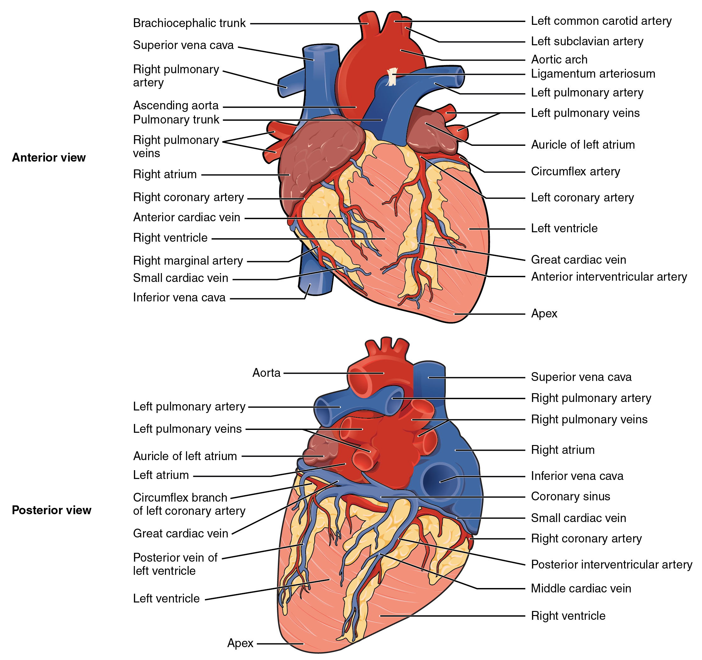
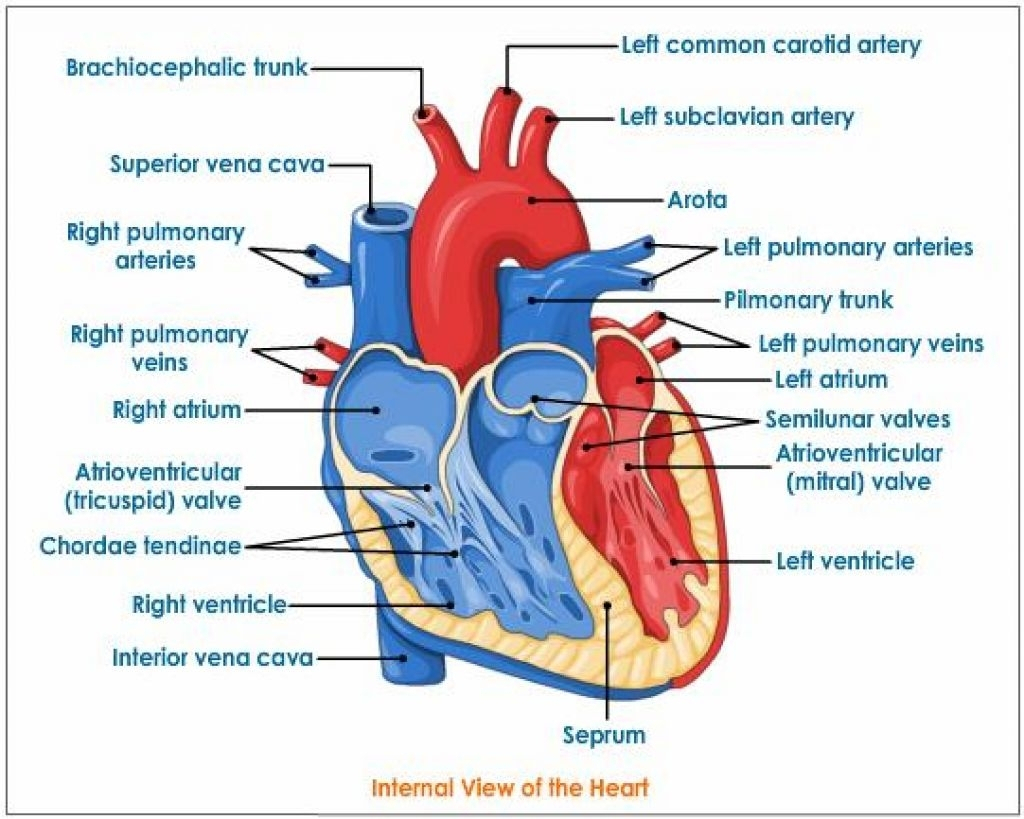
Labeled Heart Drawing - The test mode allows instant evaluation of user progress. Human anatomy diagrams show internal organs, cells, systems, conditions, symptoms and sickness information and/or tips for healthy living. Web heart, organ that serves as a pump to circulate the blood. Learn more about the heart in this article. Demarcating the area for drawing on the page. These valves have been clearly shown in the labeled diagram of the heart. Light sketching of the heart. Controls the rhythm and speed of your heart rate. [right atrium and ventricle of the heart (labeled)] Neatly print the names around your drawing and then use a ruler to draw an arrow to the corresponding part. Images are labelled, providing an invaluable medical and anatomical tool. The test mode allows instant evaluation of user progress. Identify the tissue layers of the heart; The left and right sides of the heart have different functions: Web your heart’s main function is to move blood throughout your body. Compare systemic circulation to pulmonary circulation; Identify the veins and arteries of. The left and right sides of the heart have different functions: Rotate the 3d model to see how the heart's valves control blood flow between heart chambers and blood flow out of the heart. Human anatomy diagrams show internal organs, cells, systems, conditions, symptoms and sickness information and/or. Relate the structure of the heart to its function as a pump; Compare systemic circulation to pulmonary circulation; Explore the diagram of heart along with the structural details only at byju’s. Neatly print the names around your drawing and then use a ruler to draw an arrow to the corresponding part. Web the main artery carrying oxygenated blood to all. Web the cardiovascular system. Compare systemic circulation to pulmonary circulation; Dr matt & dr mike. Human anatomy diagrams show internal organs, cells, systems, conditions, symptoms and sickness information and/or tips for healthy living. Web the heart is made of three layers of tissue. You can also refer to the byju’s app for further reference. Myocardium is the thick middle layer of muscle that allows your heart chambers to contract and relax to pump blood to your body. The heart is a hollow, muscular organ that pumps oxygenated blood throughout the body and deoxygenated blood to the lungs. Learn more about the heart in this article. Web label the urinary tract #1 printout. It consists of four chambers, four valves, two main arteries (the coronary arteries), and the conduction system. In this interactive, you can label parts of the human heart. Web describe the internal and external anatomy of the heart; Endocardium is the thin inner lining of the heart chambers and also forms the surface of the valves. Drag and drop the text labels onto the boxes next to the diagram. Web 1.3m views 3 years ago 3 products.
19.1 Heart Anatomy Anatomy and Physiology

How to Draw the Internal Structure of the Heart 14 Steps

Heart And Labels Drawing at GetDrawings Free download
If You're Trying To Identify Parts Of The Heart For A Class Or Just For Fun, Consider Adding The Names Of Each Segment.
Web Heart, Organ That Serves As A Pump To Circulate The Blood.
The Test Mode Allows Instant Evaluation Of User Progress.
Demarcating The Area For Drawing On The Page.
Related Post: