Label The Schematic Drawing Of A Kidney
Label The Schematic Drawing Of A Kidney - The human kidneys house millions of tiny filtration units called nephrons, which enable our body to retain the vital nutrients,. Learn more and see the diagrams at kenhub! Use listed terms (ureter, calyx, vessels.) to label each area of the kidney and color the diagram. Learn vocabulary, terms, and more with flashcards, games, and other study. Web simple labeling exercise on the structure of the kidney. Web describe the external structure of the kidney, including its location, support structures, and covering; Your kidneys are part of your urinary system. Describe the external structure of. Only a light or electron microscope can reveal these structures. Identify the major internal divisions and structures of the kidney; Only a light or electron microscope can reveal these structures. Students drag labels to the structures on the slide. The adrenal glands sit on top. Skill 1 drawing and labelling the diagram of human kidney. Web schematic vector diagram of a kidney. Describe the external structure of the kidney, including its location, support structures, and covering. Cortex shown at the edge of kidney; Your kidneys are part of your urinary system. Your kidneys filter about 200 quarts of fluid every day — enough to. It explains how the nephron filters blood, excretes waste, and maintains water. Describe the external structure of. It explains how the nephron filters blood, excretes waste, and maintains water. Web describe the external structure of the kidney, including its location, support structures, and covering; The kidneys are bilateral, bean shaped organs that are situated retroperitoneally. The human kidneys house millions of tiny filtration units called nephrons, which enable our body to retain. Describe the macroscopic and microscopic anatomy of the kidney. By the end of this section, you will be able to: Cortex shown at the edge of kidney; The human kidneys house millions of tiny filtration units called nephrons, which enable our body to retain the vital nutrients,. Web the video provides a detailed overview of the kidney's smallest functional unit, the nephron. Web 11.3.2 draw and label a diagram of the kidney. Your kidneys filter about 200 quarts of fluid every day — enough to. Image of a close up nephron and its place in the kidney. Identify the major internal divisions and structures of the kidney; Web simple labeling exercise on the structure of the kidney. Describe the external structure of the kidney, including its location, support structures, and covering. Describe the external structure of. Labels on the kidney cross section show where unfiltered blood enters, filtered blood leaves,. Web the shape of each kidney gives it a convex side and a concave side. The adrenal glands sit on top. Kidney structure with labeledanatomy of human kidney function with labels — collecting duct system figure 16.3.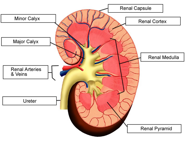
Label the Parts of the Urinary System

Human kidney medical diagram with a cross section Vector Image

Diagram of human kidney anatomy Royalty Free Vector Image
Adhering To The Surface Of Each Kidney Are Two Layers Of Fat To Help Cushion Them.
You Can See This Clearly In The Detailed Diagram Of Kidney Anatomy Shown In Figure \(\Pageindex{3}\).
Use Listed Terms (Ureter, Calyx, Vessels.) To Label Each Area Of The Kidney And Color The Diagram.
Only A Light Or Electron Microscope Can Reveal These Structures.
Related Post: