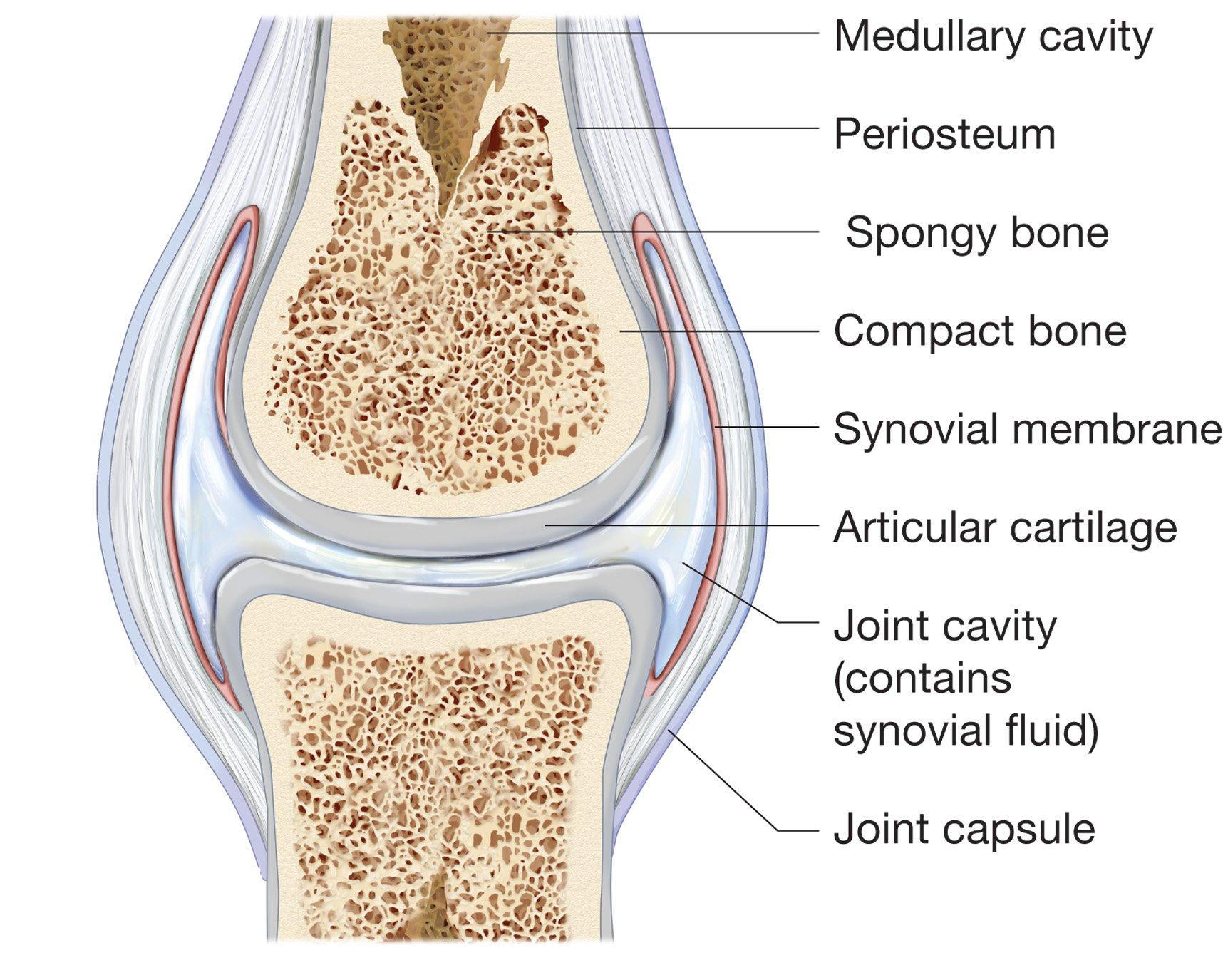
Joint Drawing Biology
Joint Drawing Biology - Web a synovial joint is a connection between two bones consisting of a cartilage lined cavity filled with fluid, which is known as a diarthrosis joint. In this chapter, we describe major components of synovium and derived tissues, including synovial membrane, synovial cells, and synovial fluid. Web describe the structural features of a synovial joint. Compare the six types of synovial joints; The humerus of the arm and the radius and the ulna of the forearm. Watch the following videos for a tour of the ankle, knee, hip, shoulder, and elbow joints. It consists of several components which can be labelled on a diagram and annotated with descriptions of each structure in the following way: Structures of the elbow joint. Explain the role of joints in skeletal movement. List the six types of synovial joints and give an example of each. Classify the different types of joints on the basis of structure. Web describe the structures that support and prevent excess movements at each joint. Begin by optimizing the flow of information (8:11). Cut it off, so that you have a roughly circular area with the skin removed. Each synovial joint of the body is specialized to perform certain movements. Web describe the structural features and functional properties of a synovial joint; Anatomy and physiology i (lumen) 12: Drawing biological pathways is complex and intimidating (2:03), but shiz shares her expertise in scientific illustration to help you start illustrating better figures today with 4 tips (5:11). Structures of the elbow joint. It consists of several components which can be labelled. Web you will need to cut about 1 mm deep. Discuss the function of additional structures associated with synovial joints; Describe the three functional types of joints and give an example of each. Explain the role of joints in skeletal movement. You can see a drawing of a typical synovial joint in figure 14.6.2 14.6. Watching these videos will help you better visualize the way joints work in the body. You can see a drawing of a typical synovial joint in figure 14.6.2 14.6. Web sir benjamin collins brodie, 1st baronet. Locomotion is the ability to move from one place to another. Distinguish between the functional and structural classifications for joints. Classify the different types of joints on the basis of structure. Describe the three functional types of joints and give an example of each. Synovial joints are further classified into six different categories on the basis of the shape and structure of the joint. The elbow is the joint connecting the upper arm to the forearm. Identify the six types of synovial joints. The hinge joint is one of six types of synovial joints along with the plane, ellipsoid, ball and socket, pivot and saddle joints. By the end of this section, you will be able to: Discuss the function of additional structures associated with synovial joints; Synovial joints are the most common types of joints in the human body. Describe the characteristic features for fibrous, cartilaginous, and synovial joints and give examples of each. Each synovial joint of the body is specialized to perform certain movements.
Classification of Joints Online Biology Notes

How to draw a synovial joint easily YouTube

Drag The Labels Onto The Diagram To Identify The Structures And
Discuss Both Functional And Structural Classifications For Body Joints.
Describe The Structural Features Of A Synovial Joint.
Web Jeff Driban, Ann E.
This Chapter Covers Many Aspects Of Joint Anatomy, Histology, And Cell Biology.
Related Post: