Homunculus Drawing
Homunculus Drawing - Web to construct a model of the homunculus that is a map of your own somatosensory cortex. The human brain’s motor cortex is often regarded as a linear map with discrete sections, each controlling different parts of the body. The pink and brown blobs are its whiskers. Web the establishment of the homunculus, a schematic drawing reflecting the disproportional representation of the parts of the human body on the motor and somatosensory cortex, was an important milestone for the neurosciences. This is a technique that neurologists commonly use on patients to diagnose nerve injury. Web textbook homunculus diagram depicts how the brain controls individual body parts — the revamp could improve treatments for brain injury. The homunculus or ‘little man’ is a. Rebekah corlew and theo walker of the fitzpatrick lab at the max planck florida for neuroscience in jupiter. Web this odd map, corresponding body part to touch sensitivity, is called a homunculus, latin for little man. Motor and sensory representation of the body in the cortex. Web textbook homunculus diagram depicts how the brain controls individual body parts — the revamp could improve treatments for brain injury. Web the shape of a mouse's body as represented in its brain. The classical view of how the human brain controls voluntary movement might not tell the whole story. Rebekah corlew and theo walker of the fitzpatrick lab at. Web the shape of a mouse's body as represented in its brain. The drawing shows some areas of the brain associated with different parts of the body. Web march 16, 2015 at 1:22 pm. There is one of these maps for our movements. It is a subjective test, requiring the Web view the region in different wavelengths of light, fly through a 3d visualization of the star system, and 3d print your own homunculus nebula. Click here to start exploring. This experiment was developed as a neuroscience outreach tool by dr. Watch the video tutorial now. Motor and sensory representation of the body in the cortex. Web textbook homunculus diagram depicts how the brain controls individual body parts — the revamp could improve treatments for brain injury. This drawing shows that areas of your body are represented disproportionately in the somatosensory cortex, and was discovered in 1950 by canadian neurosurgeon dr. Watch the video tutorial now. The drawing shows some areas of the brain associated with different parts of the body. It is a subjective test, requiring the Web the homunculus is commonly used today in scientific disciplines such as psychology as a teaching or memory tool to describe the distorted scale model of a human drawn or sculpted to reflect the relative space human body parts occupy on the somatosensory cortex (the sensory homunculus) and the motor cortex (the motor homunculus). Web map your own brain in 10 minutes or less. It has wide eyes, huge hands and feet weighing down like rocks, and a swollen tongue sticking out through an. Body part pictures of various sizes, taped together: You know what you look like, but what does your brain think you look like? That map of the primary motor cortex — the motor. Web the establishment of the homunculus, a schematic drawing reflecting the disproportional representation of the parts of the human body on the motor and somatosensory cortex, was an important milestone for the neurosciences. This is a technique that neurologists commonly use on patients to diagnose nerve injury. Nature reviews molecular cell biology 13 , 410 ( 2012) cite this article. Nature neuroscience, august 2013, doi:10.1038/nn.3454. Web to construct a model of the homunculus that is a map of your own somatosensory cortex.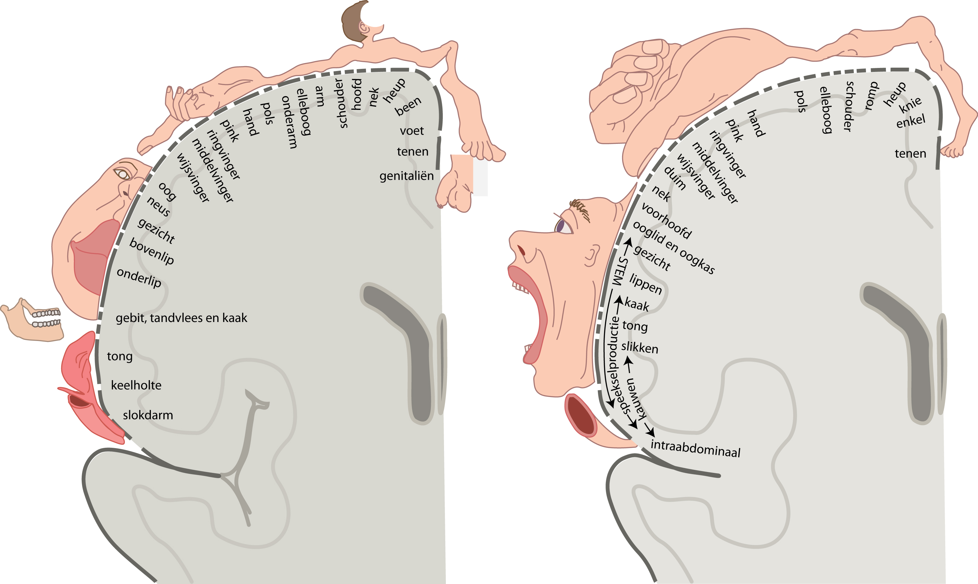
Slagter Drawing Cortical motor and sensory homunculus Dutch labels

Cortical Homunculus Illustration Stock Image C043/2707 Science
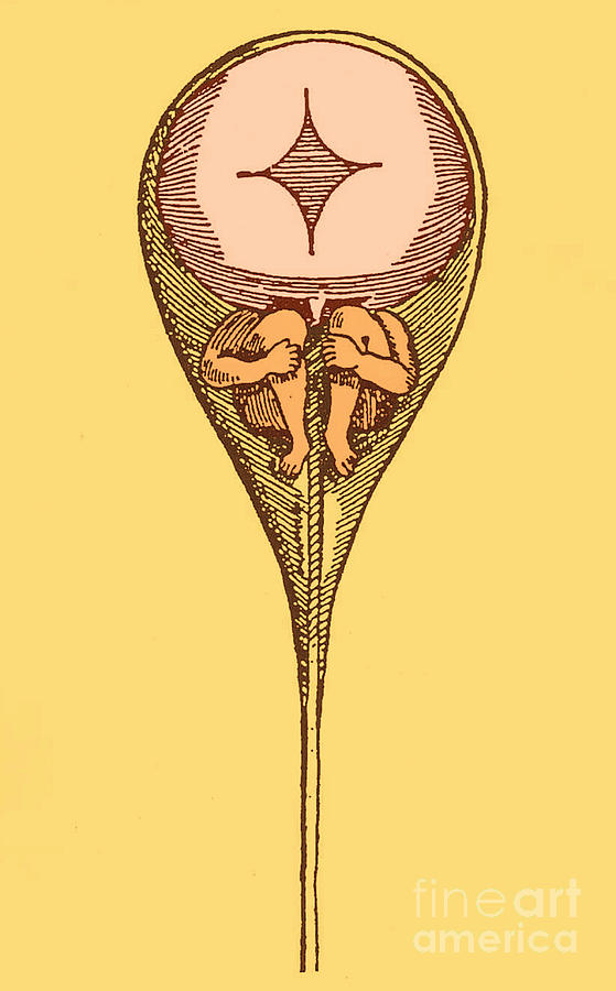
Drawing Of Homunculus, 1694 Photograph by Science Source Pixels
Web Revealing The Missing Female Homunculus.
Eighty Years Ago, Penfield And Boldrey Electrically Stimulated The Cortical Surface Of Patients Undergoing.
The Pink And Brown Blobs Are Its Whiskers.
Web The Neurological Homunculus Was Generated From 170 Summary Maps Of The Number And Location Of Stimulation Points For Each Body Part, Each Patiently Sketched By Boldrey From Penfield’s Operation Notes, Photographs And Drawings.
Related Post: