Heart Drawing Labeled
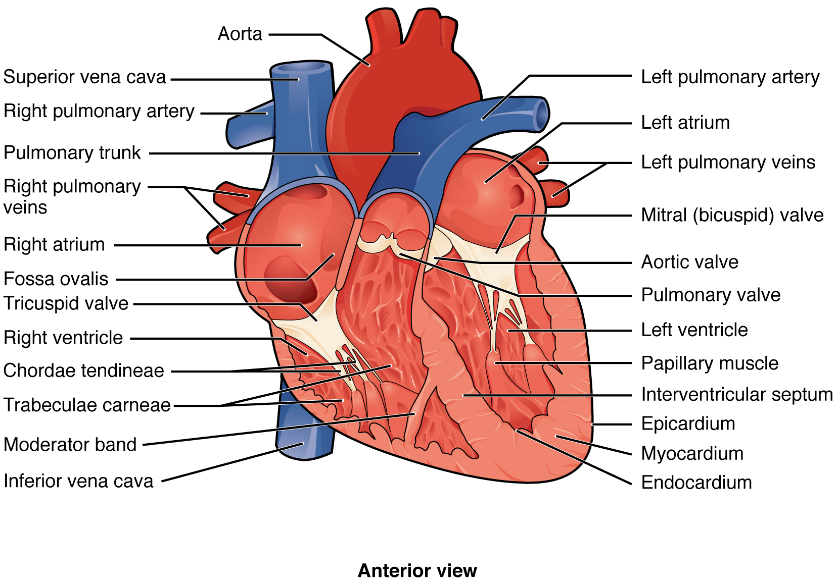
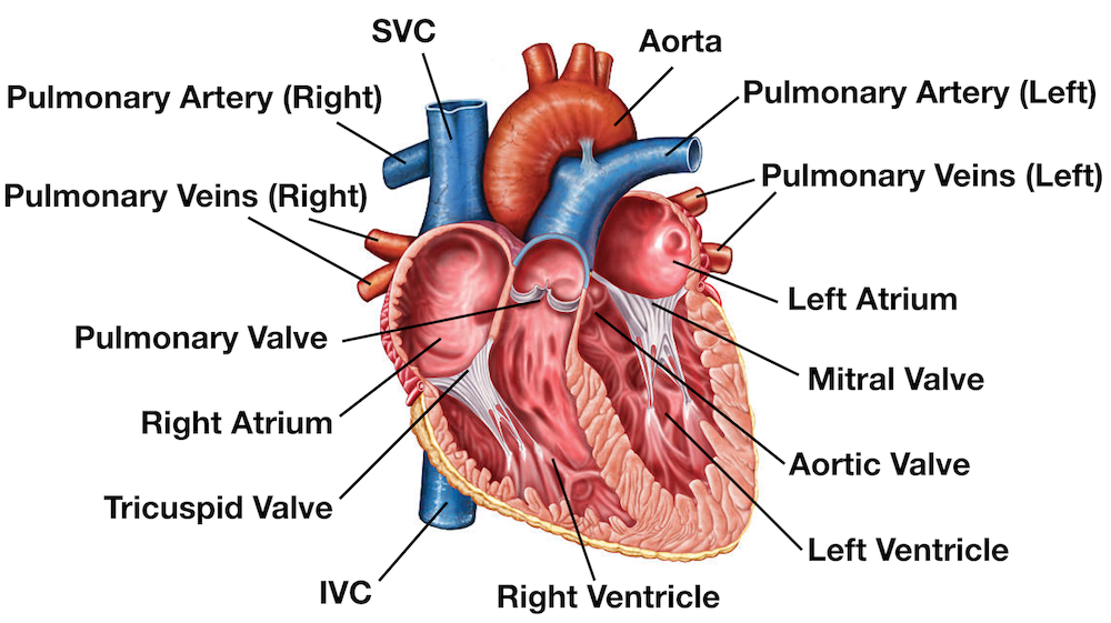
Heart Drawing Labeled - The heart is divided into four chambers: Web your heart’s main function is to move blood throughout your body. Compare systemic circulation to pulmonary circulation. Anatomy of the heart made easy along with the blood flow through the cardiac structures, valves, atria, and ventricles. Trace the pathway of oxygenated and deoxygenated blood thorough the chambers of the heart. This tool provides access to several medical illustrations, allowing the user to interactively discover heart anatomy. Find an image that displays the entire heart, and click on it to enlarge it. New 3d rotate and zoom. Web dr matt & dr mike. The heart is a hollow, muscular organ that pumps oxygenated blood throughout the body and deoxygenated blood to the lungs. Anatomy of the heart made easy along with the blood flow through the cardiac structures, valves, atria, and ventricles. Once you’re feeling confident, you can test yourself using the unlabeled diagrams of the parts of the heart below. Identify the veins and arteries of. How to visualize anatomic structures. The two upper chambers are called the left and the right. Controls the rhythm and speed of your heart rate. [right atrium and ventricle of the heart (labeled)] Identify the tissue layers of the heart; The heart is divided into four chambers: Web in just a few minutes, you will be able to label the entire diagram shown below! This tool provides access to several medical illustrations, allowing the user to interactively discover heart anatomy. Web your heart’s main function is to move blood throughout your body. Web the human heart, comprises four chambers: Labeled heart diagram showing the heart from anterior. Web in just a few minutes, you will be able to label the entire diagram shown below! Identify the veins and arteries of. Web inside, the heart is divided into four heart chambers: It’s nearly the size of an adult’s clenched fist. Web label the heart. Controls the rhythm and speed of your heart rate. Blood is transported through the body via a complex network of veins. The two atria and ventricles are separated from each other by a muscle wall called ‘septum’. Neatly print the names around your drawing and then use a ruler to draw an arrow to the corresponding part. Web function and anatomy of the heart made easy using labeled diagrams of cardiac structures and blood flow through the atria, ventricles, valves, aorta, pulmonary arteries veins, superior inferior vena cava, and chambers. Blood brings oxygen and nutrients to your cells. In this interactive, you can label parts of the human heart. The video above also provides an animation at the end to quiz yourself and test your knowledge! Start with the pulmonary veins. Your heart is located between your lungs in the middle of your chest, behind and slightly to the left of your breastbone. Images are labelled, providing an invaluable medical and anatomical tool. Web in just a few minutes, you will be able to label the entire diagram shown below!
OpenStax AnatPhys fig.19.9 Internal Anatomy of the Heart English
Heart Anatomy Labeled Diagram, Structures, Blood Flow, Function of
.svg/1043px-Diagram_of_the_human_heart_(cropped).svg.png)
FileDiagram of the human heart (cropped).svg Wikipedia
Web © 2024 Visible Body.
Web 1.3M Views 3 Years Ago 3 Products.
Compare Systemic Circulation To Pulmonary Circulation.
Difference Between Arteries And Veins.
Related Post:
