Ependymal Cells Drawing
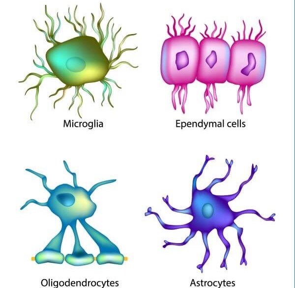
Ependymal Cells Drawing - Ependymal cells in lateral ventricles (lv) are mainly e1 types, while e1, e2, e3 cell in v3 (1). Web ependymal cells are one of the four main types of glial cells, and themselves encompass three types of cells 1: Multiciliated ependymal cells line all brain cavities. The beating of their motile cilia contributes to the flow of cerebrospinal fluid, which. Microglia are a type of glial cell which are essential for the defense mechanisms in the central nervous system (cns). 4 three specific subtypes of ependymal cells h. They are indispensable components of the central nervous system (cns) and originate from neuroepithelial cells of the neural plate. Web photomicrograph of the ependymal cell lining stained with haematoxylin and eosin. These cells line the ventricles in the brain and the central canal of the spinal cord, which become filled with cerebrospinal fluid. They play a critical role in cerebrospinal fluid (csf) homeostasis, brain metabolism, and. Web the ependyma is made up of ependymal cells called ependymocytes, a type of glial cell. These cells line the ventricles in the brain and the central canal of the spinal cord, which become filled with cerebrospinal fluid. Web this video describes the structure and function of ependymal cells. Web ependymal cells ependymal cells, which create cerebral spinal fluid (csf),. There is no definition for this structure yet. Microglia share structural and functional similarities with tissue macrophages, highlighting their role in immune responses within the cns. Microglia are a type of glial cell which are essential for the defense mechanisms in the central nervous system (cns). Line the floor of the third ventricle overlying the median eminence of the hypothalamus. Microglia are a type of glial cell which are essential for the defense mechanisms in the central nervous system (cns). How do you know where you are right now? They are indispensable components of the central nervous system (cns) and originate from neuroepithelial cells of the neural plate. Web ependymocytes are one of the three types of ependymal cells, which. Web ependymal cells ependymal cells, which create cerebral spinal fluid (csf), line the ventricles of the brain and central canal of the spinal cord. Structure of mouse ventricle (top left) from the allen brain atlas with different ependymal cell subtypes in distinct ventricular regions (1) (2) (3). Suggest a definition i agree herein to the cession of rights to my contribution in accordance with the terms and conditions of the website. How do you know where you are right now? They are indispensable components of the central nervous system (cns) and originate from neuroepithelial cells of the neural plate. Web ependymocytes are one of the three types of ependymal cells, which in turn are one of the four principles types of glial cells, and are found lining the ventricular system of the brain and the central canal of the spinal cord 1. Their main function is to secrete, circulate, and maintain homeostasis of the cerebrospinal fluid that fills the ventricles of the central nervous system. Scale bar represents 70 µm. The beating of their motile cilia contributes to the flow of cerebrospinal fluid, which. Web schematic diagram of the morphology and distribution of ependymal cells. Ependymal cells also give rise to the epithelial layer that surrounds the choroid plexus, a network of blood vessels located. These cells line the ventricles in the brain and the central canal of the spinal cord, which become filled with cerebrospinal fluid. Line the ventricles of the brain and central canal of the spinal cord; Line the floor of the third ventricle overlying the median eminence of the hypothalamus Web the neuroepithelium and ependyma constitute barriers containing polarized cells covering the embryonic or mature brain ventricles, respectively; In addition, ependymal cells are suggested to be latent nscs with a capacity to acquire neurogenic function.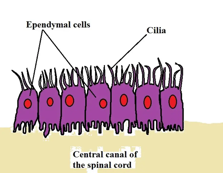
Ependymal Cells Diagram
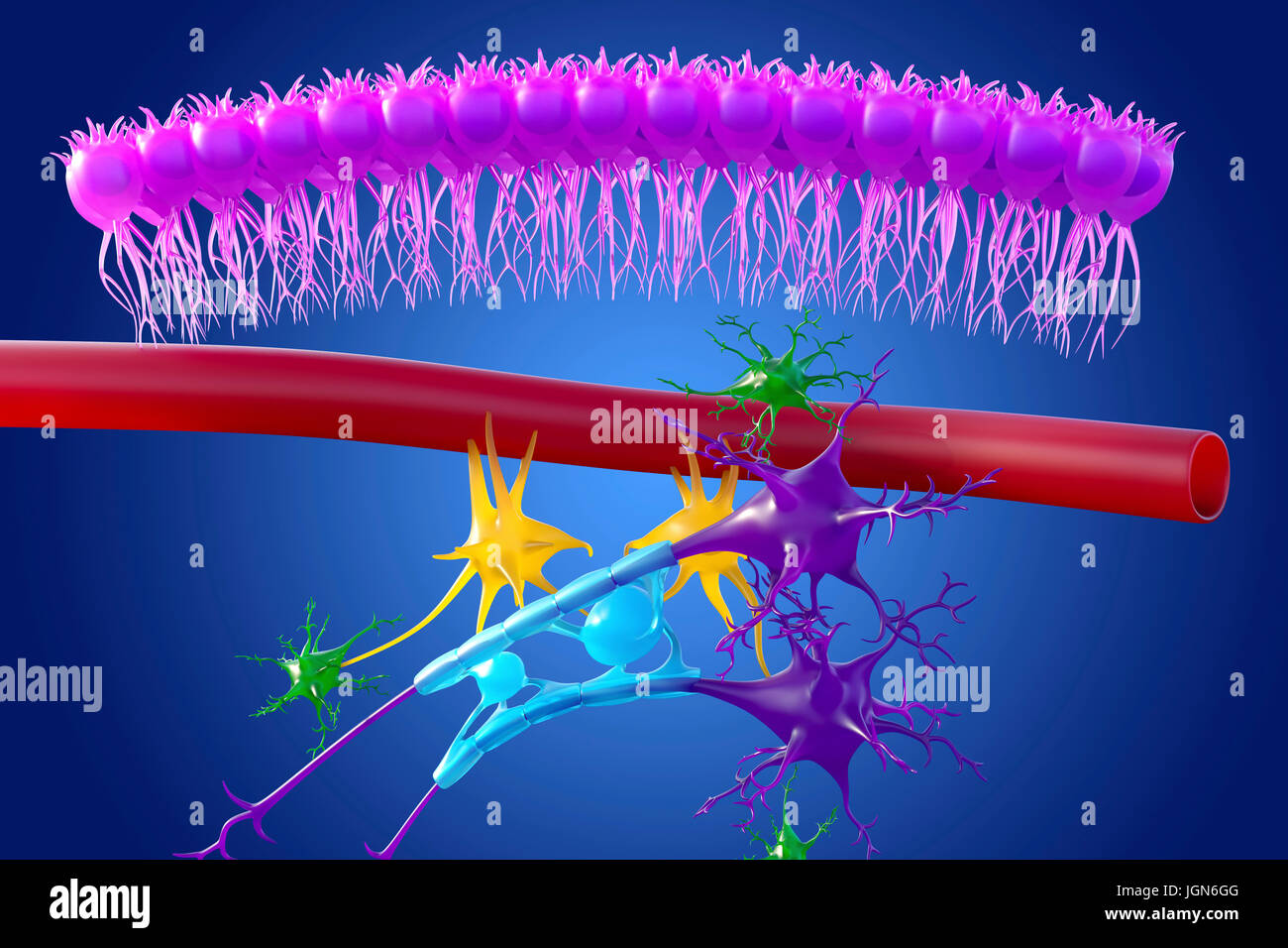
Brain nervous tissue, illustration. Seen here are ependymal cells (pink
Ependymal Cell The Definitive Guide Biology Dictionary
Introduction To Neurons And Glia.
Therefore, They Separate The Cerebrospinal Fluid That Fills Cavities From.
The Standard Morphological Features Of Ependymal Cells Include A Large Oval Nucleus, Short Microvilli, And Long Cilia That Project Outwards Into The Ventricular Space.
They Play A Critical Role In Cerebrospinal Fluid (Csf) Homeostasis, Brain Metabolism, And.
Related Post: