Drawing Of The Uterus
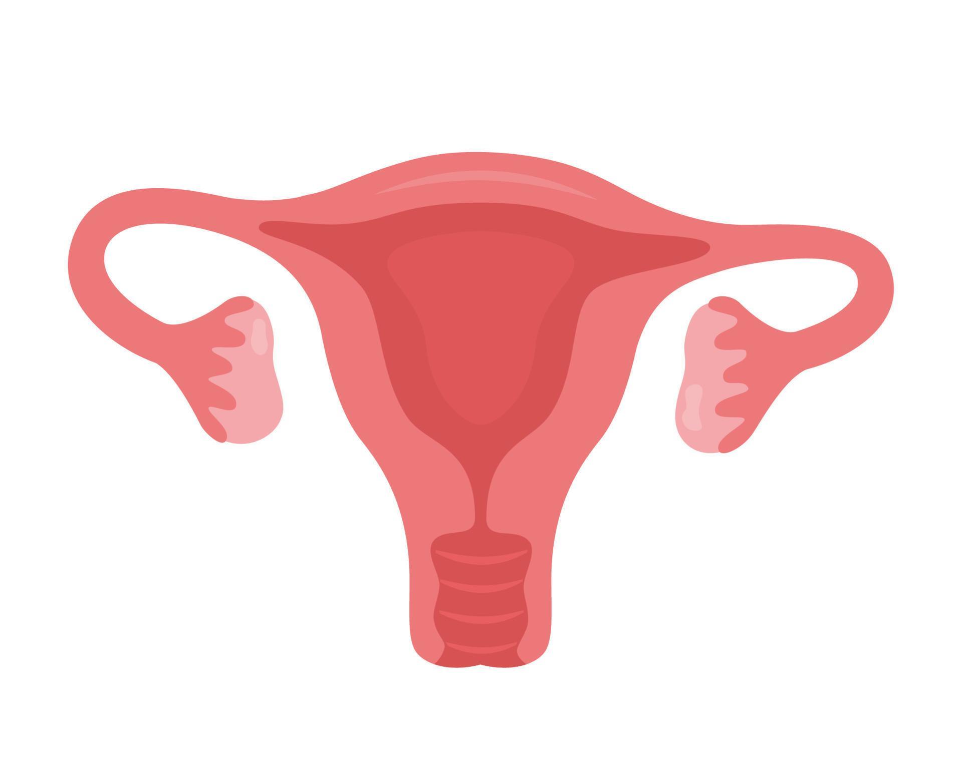
Drawing Of The Uterus - In this sagittal view of the female pelvic cavity, identify the primary reproductive organ and the accessory reproductive organs shown. The uterus is where a fetus develops during pregnancy. Illustrative drawings in detail of the placenta and. This short article describes the normal anatomy of the uterus and will focus on definitions, structure, location, supporting ligaments, blood supply and innervation. Web how to draw uterus easily | female reproductive system @justmadeeasyto save your time and excel your knowledge, i post related video links in the discription. Web the uterus has three parts; In the lower study leonardo removes the uterine wall to show the calf; Browse 668 uterus drawing photos and images available, or search for uterus illustration to find more great photos and pictures. It's also called the womb. The uterus, also known as the womb, is a hollow, muscular organ located in the pelvis between the bladder and rectum. Web the uterus is the muscular organ that nourishes and supports the growing embryo (see figure 27.14). A large drawing of an embryo within a human uterus with a cow's placenta; Browse 668 uterus drawing photos and images available, or search for uterus illustration to find more great photos and pictures. This part is structurally and functionally different to the. Web label the wall of the uterus using the hints provided. Web the uterus is the muscular organ that nourishes and supports the growing embryo (see figure 27.14). In this sagittal view of the female pelvic cavity, identify the primary reproductive organ and the accessory reproductive organs shown. The placenta is represented as a pattern of small ovals. The uterus. However, about 4% of females have a uterus that has a different shape. It is usually present in people assigned female at birth. This short article describes the normal anatomy of the uterus and will focus on definitions, structure, location, supporting ligaments, blood supply and innervation. Web the uterus is the muscular organ that nourishes and supports the growing embryo. Web anatomy of the female reproductive system; Drawing shows the uterus, myometrium (muscular outer layer of the uterus), endometrium (inner lining of the uterus), ovaries, fallopian tubes, cervix, and vagina. This part is structurally and functionally different to the rest of the uterus. Illustrative drawings in detail of the placenta and. Web the uterus is the muscular organ that nourishes and supports the growing embryo (see figure 27.14). The uterus has three layers: However, about 4% of females have a uterus that has a different shape. Web label the wall of the uterus using the hints provided. The female reproductive organs include several key structures, such as the ovaries, uterus, vagina, and vulva. The placenta is represented as a pattern of small ovals. Web the uterus (from latin uterus, pl.: Its average size is approximately 5 cm wide by 7 cm long (approximately 2 in by 3 in) when a female is not pregnant. This system of ducts connects to the ovaries, the primary reproductive organs. The function of the uterine cervix during pregnancy is described at the end of the article. These fully annotated anatomical illustrations are presented as a comprehensive atlas of the female reproductive system, bladder, rectum and. The endometrium (uterine mucous membrane) is lined with simple columnar epithelium (lamina epithelialis) and contains numerous tubular glands.![]()
Vector Isolated Illustration of Uterus Stock Vector Illustration of
![]()
This Icon Represents The Uterus Of A Female Human Uterus Png Clipart
Uterus. Woman reproductive health illustration. Gynecology. Anatomy
It's Also Called The Womb.
Mucosa (Endometrium), Muscularis ( Myometrium) And Serosa / Adventitia ( Perimetrium ).
A Smaller Sketch Of The Same;
In This Sagittal View Of The Female Pelvic Cavity, Identify The Primary Reproductive Organ And The Accessory Reproductive Organs Shown.
Related Post: