Drawing Of Simple Squamous Epithelium
Drawing Of Simple Squamous Epithelium - Simple epithelium can be divided into 4 major classes, depending on the shapes of constituent cells. Use the image slider below to learn how to use a microscope to identify and study simple squamous epithelium in renal corpuscles of the renal (kidney) cortex. 19k views 2 years ago cell biology. Web there are three basic shapes used to classify epithelial cells. Histology of the simple squamous epithelium that lines the renal corpuscles in the kidney. A cuboidal epithelial cell looks close to a square. A columnar epithelial cell looks like a column or a tall rectangle. This epithelium presents a minimal barrier to passive diffusion and, therefore, lines surfaces across which metabolites or gases can move rapidly. The typical example of the simple squamous epithelium will be found in the lung’s alveoli, the parietal layer of the bowman’s capsule of the kidney, and the loop of henle of kidney tubules. The simple squamous epithelium consists of cells that are thinly walled with a dense nucleus. 2.6k views 3 years ago pakistan. Each epithelial tissue has a top (apical) surface, a bottom (basal) surface and side (lateral) surfaces. Web structure of the simple squamous epithelium. Web what is simple squamous epithelium? Use the image slider below to learn more about the characteristics of stratified squamous epithelium. Web drawing histological diagram of simple squamous epithelia.useful for all medical students.drawn by using h & e pencils Web a simple squamous epithelium, also known as pavement epithelium and tessellated epithelium, is a single layer of flattened, polygonal cells in contact with the basal lamina (one of the two layers of the basement membrane) of the epithelium. 2.6k views 3. Web simple epithelial tissue is made up of a single layer of cells, attached to a layer of connective tissue called the basement membrane. Use the image slider below to learn more about the characteristics of stratified squamous epithelium. Web clark brelje and robert l. Describe the structure and function of endocrine and exocrine glands. Simple squamous epithelium, isolated (400x). Squamous cells are large, thin, and flat and contain a rounded nucleus. Use the image slider below to learn more about the characteristics of simple squamous epithelium. Like other epithelial cells, they have polarity and contain a distinct apical surface with specialized membrane proteins. The cells are tightly packed together due to pressure, giving them a polygonal arrangement. This epithelium presents a minimal barrier to passive diffusion and, therefore, lines surfaces across which metabolites or gases can move rapidly. Web simple squamous epithelium can be found in many locations in the body (e.g., lining blood vessels, lining the alveoli (air sacs) of our lungs, and in bowman’s capsule of the kidney). Web drawing histological diagram of simple squamous epithelia.useful for all medical students.drawn by using h & e pencils Both the endothelial lining of blood vessels and the mesothelial lining of the body cavities are simple squamous epithelium. Web simple squamous epithelium, isolated (40x) buccal mucosal. Each epithelial tissue has a top (apical) surface, a bottom (basal) surface and side (lateral) surfaces. Web there are three basic shapes used to classify epithelial cells. • how to draw simple squamous epitheliu. Compare this image to the drawing of simple squamous epithelium in your textbook, which shows what an intact layer of cells should look like. Web simple squamous epithelium is composed of a single layer of thin cells that are much wider than they are tall. Simple squamous epithelium is thin and flat. The position of the nucleus depends on the form of the cells where the nucleus is mostly randomly oriented towards the periphery.
Simple Squamous Epithelium Diagram Quizlet

How to draw stratified squamous epithelium easy way YouTube
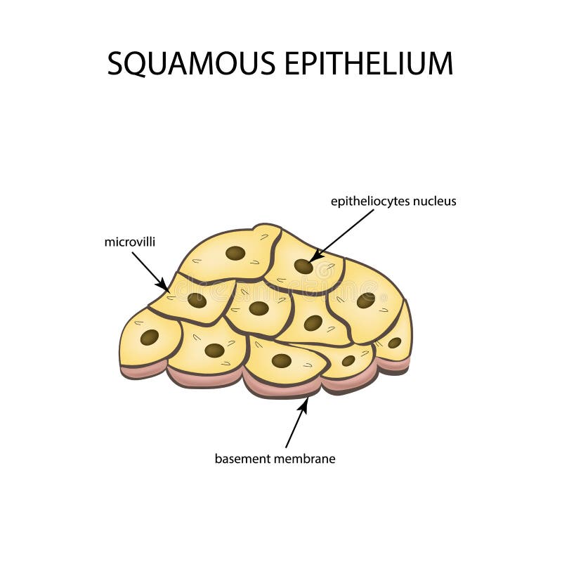
The Structure of the Squamous Epithelium. Infographics. Vector
Web In This Portion, I Will Show You The Simple Squamous Epithelium Labeled Diagrams From The Different Organs Or Parts, Or Structures Of The Animal’s Body.
Web Structure Of The Simple Squamous Epithelium.
Web Distinguish Between Simple Epithelia And Stratified Epithelia, As Well As Between Squamous, Cuboidal, And Columnar Epithelia.
A Squamous Epithelial Cell Looks Flat Under A Microscope.
Related Post: