Drawing Of Knee Anatomy
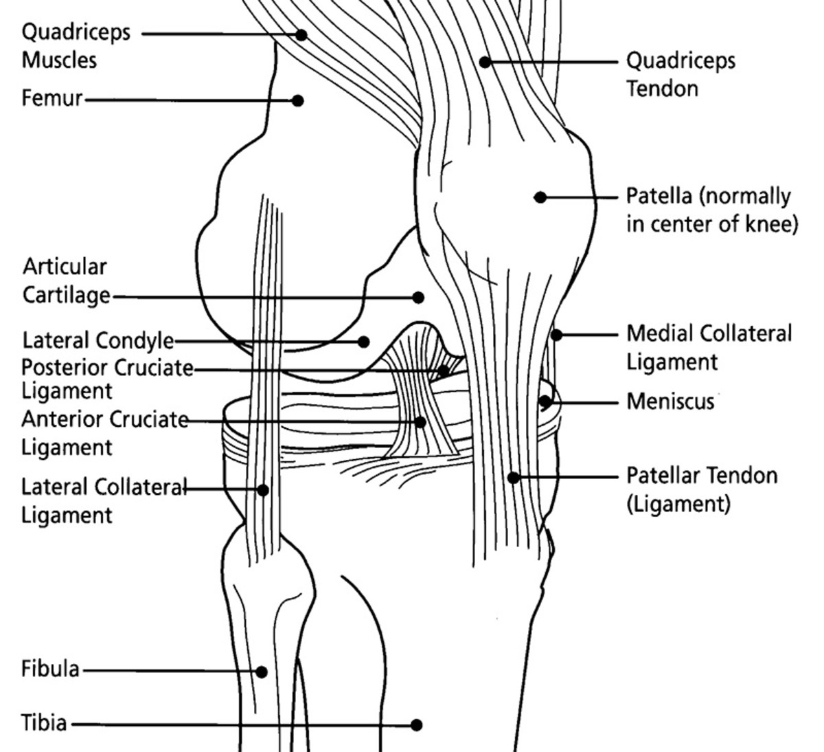
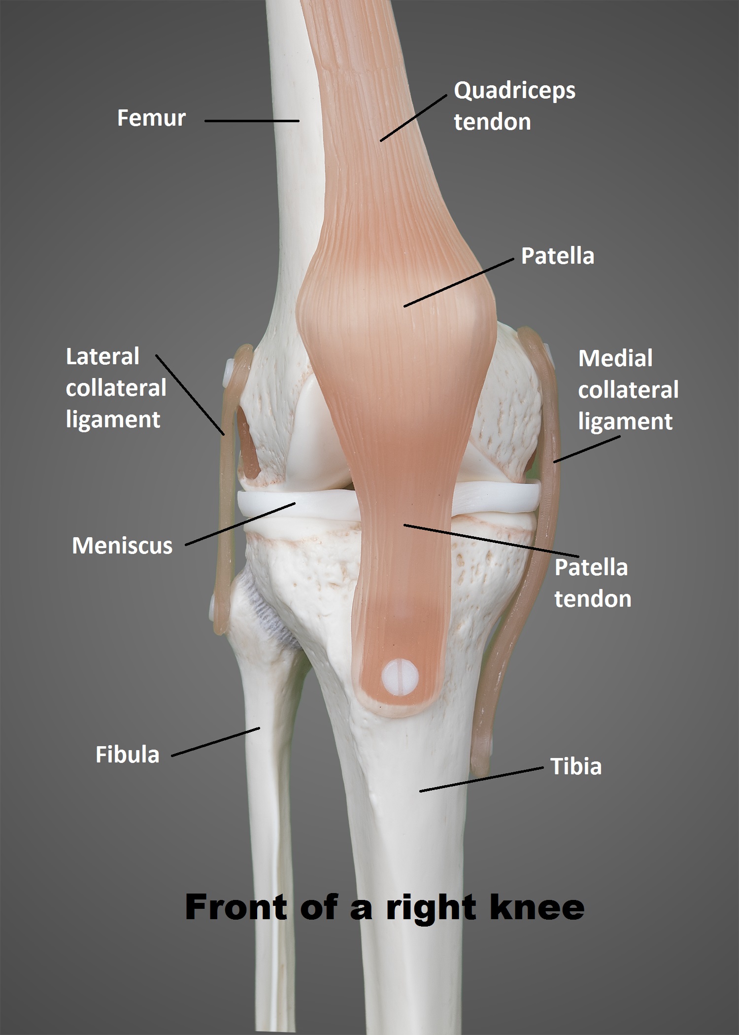
Drawing Of Knee Anatomy - Stabilizing you and helping keep your balance. Which ligaments keep it stable? The knee joint is one of the largest and most complex joints in the body. Web the knee joint is the largest joint in the body and connects the thigh with the lower leg. Web the femur (thigh bone), tibia (shin bone), and patella (kneecap) make up the bones of the knee. Where is the knee joint located? Learning about knee anatomy can help the students to know about the structure and function of the knee. Web anatomy of the knee. The knee is a complex joint that flexes, extends, and twists slightly from. See possible causes of severe knee pain. Web the femur (thigh bone), tibia (shin bone), and patella (kneecap) make up the bones of the knee. Adjacent and attached to the tibia is the fibula. Click to view large image. Stabilizing you and helping keep your balance. The patella is a small, triangle shaped bone that sits. See knee drawing stock video clips. Knee anatomy involves more than just muscles and bones. Supporting your body when you stand and move. The knee is the joint in the middle of your leg. In the knee joint, the femur articulates with the tibia and the patella. The knee joint is a complex hinge joint. The knee joint is the largest joint in the human body, and the joint most commonly affected by arthritis. The kneecap itself is known as the patella. Vector human knee joint symbols. Anterior view showing the anatomy of the patella, femur, tibia, fibula and femorotibial joint. They are attached to the femur (thighbone), tibia (shinbone), and fibula (calf bone) by fibrous tissues called. The knee joint is the largest joint in the human body, and the joint most commonly affected by arthritis. By nature, your audience has a basic understanding of how the human body is supposed to look and move. Web anatomy of the knee. Web the knee joint is the largest joint in the body and connects the thigh with the lower leg. Web the main features of the knee anatomy include bones, cartilages, ligaments, tendons and muscles. The muscles that affect the knee’s movement run along the thigh and calf. Web the structure of a normal knee joint. Stabilizing you and helping keep your balance. Finally, draw in the hamstrings covering the calves at the back of the knee. Why learn how to draw human anatomy. The knee joint keeps these bones in place. The largest joint in the body, the knee is also one of the most easily injured. The kneecap itself is known as the patella. The knee joint is a complex hinge joint. Cartoon illustration of the human knee joint anatomy.
FileKnee diagram.svg Wikipedia

Anatomy of the Knee Joint (With Diagrams and XRay) Owlcation

The Knee UT Health San Antonio
Where Is The Knee Joint Located?
It Is Made Up Of Two Joints, The Tibiofemoral Joint (Between The Tibia And The Femur), And The Patellofemoral Joint (Between The Patella And The Femur).
The Upper Leg Is Facing The Viewer And Can Be Simplified As An Elongated Cuboid Shape In Perspective.
Web To Draw The Knee, Begin By Visualizing The Bones And Tendons Underneath To Help With The Placement Of Landmarks.
Related Post: