Drawing Of Bladder
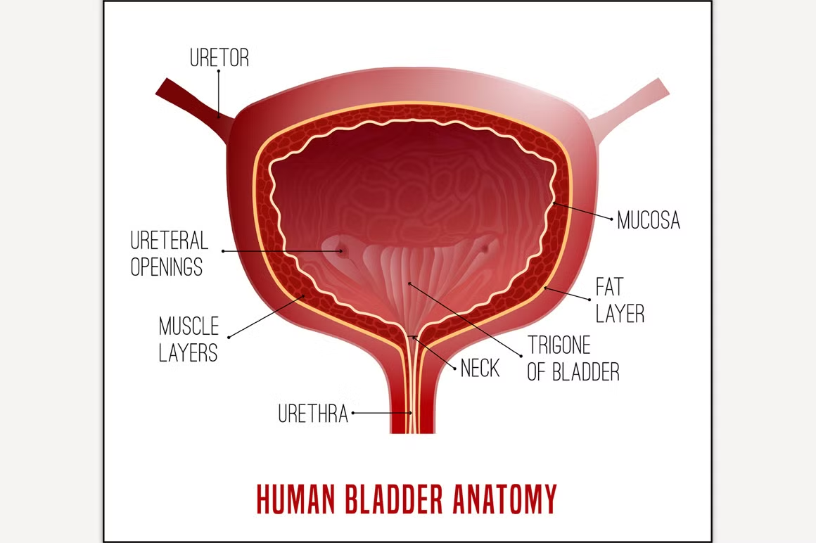
Drawing Of Bladder - Web anatomy of the female urinary system showing the kidneys, ureters, bladder, and urethra. The bladder's walls relax and expand to store urine, and contract and flatten to. 211 views 10 months ago. The inner lining of the bladder tucks into the folds and expands out to. Some messages are easier to explain. Use the menu to see other pages. You will find drawings of the bladder and its tissue layers. The body, apex, fundus and neck: Web the urinary bladder is a muscular sac whose function is to temporarily store urine. Web drawing of bladder pictures, images and stock photos. Its walls consist of smooth muscle which allows the bladder to stretch, permitting the bladder to store an increasing amount of urine. They can make urinating painful, difficult or uncontrollable. Web anatomy of the female urinary system showing the kidneys, ureters, bladder, and urethra. If you down a lot of water, it's likely that you'll need to urinate soon. It. Use the menu to choose a different section to read in this guide. It plays two main roles: Female anatomy includes the internal and external structures of the reproductive and urinary systems. Additionally, it is divided into four main parts: 211 views 10 months ago. The kidneys, ureters, bladder, and urethra are labeled. If you down a lot of water, it's likely that you'll need to urinate soon. It is held in place by ligaments that are attached to other organs and the pelvic bones. Overview of the urinary tract. 1.006 mb | 2175 x 2475. Use the menu to see other pages. Urine is made in the renal tubules and collects in the renal pelvis of each kidney. The urinary system helps rid the body of toxins through urination (peeing). It plays two main roles: The next section in this guide is risk factors. It describes the factors that may increase the chance of developing bladder cancer. Urine produced by the kidneys flows through the ureters to the urinary bladder, where is it stored before passing into the urethra and exiting the body. F t k e p. The bladder is shown in cross section to reveal interior wall and openings where the ureters empty into the bladder. 1.006 mb | 2175 x 2475. Additionally, it is divided into four main parts: Drawing of the urinary tract inside the outline of the upper half of a human body. Collection of human internal organs hand drawn sketch style. Web anatomy and function of the urinary bladder. The body, apex, fundus and neck: Most people urinate six to eight times a day, and this regular act can reveal much about your bladder's health.
Bladder Anatomy Image CustomDesigned Illustrations Creative Market

'Bladder, drawing' Stock Image C002/0616 Science Photo Library

Cross section bladder human internal organ Vector Image
Every Day, You Get Direct Feedback From A Vital Organ:
You Will Find Drawings Of The Bladder And Its Tissue Layers.
The Urinary Bladder Is A Pelvic Organ That Collects And Holds Urine Before Urination.
Female Anatomy Includes The Internal And External Structures Of The Reproductive And Urinary Systems.
Related Post: