Drawing Cranial Nerves
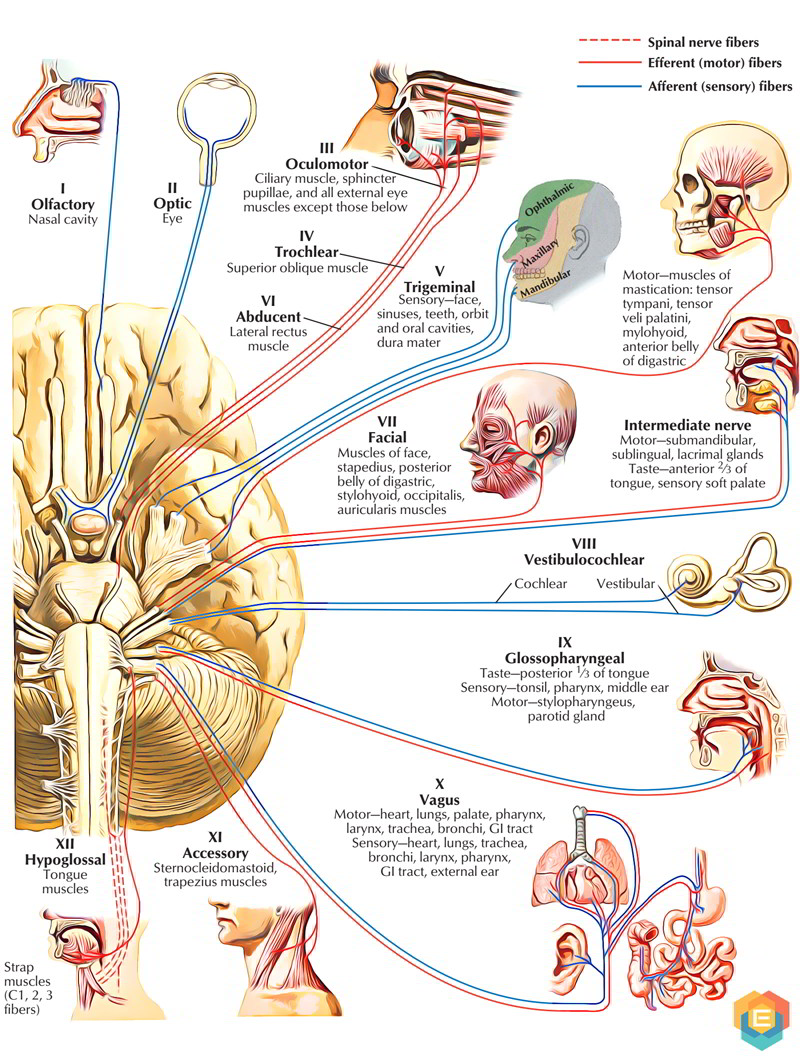
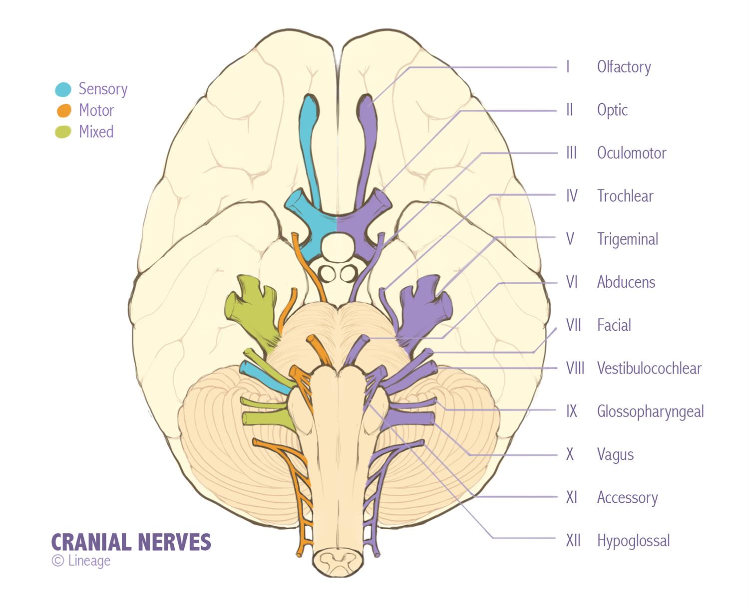
Drawing Cranial Nerves - The cranial nerves are loosely based on their functions. Web a number of cranial nerves send electrical signals between your brain and different parts of your neck, head and torso. To begin, draw the cervical spinal cord. Web the cranial nerves can be considered both in terms of their anatomical numbering from i to xii, which describes their sequential origins from the caudal to the ventral brainstem, or in groups according to their. Central nerves are in your brain and spinal cord. The nervous system is divided into two main parts: Nasal mucous membrane high in the nasal cavities. Web cranial nerves , anatomy : These original anatomical drawings were produced digitally, working from medical imaging sources and 3d reconstructions using adobe illustrator. In this summary, we discuss the nomenclature of the cranial nerves and supply some background information that might make it easier to understand the nerves and their function. Web magnetic resonance imaging (mri) is the gold standard technique in the study of the cranial nerves. By the end of this section, you will be able to: Name the twelve cranial nerves and explain the functions associated with each. All cranial nerves originate from nuclei in the brain. Let's start with an anterior view of the brainstem, which is. Web magnetic resonance imaging (mri) is the gold standard technique in the study of the cranial nerves. In this video i will go over cranial nerves i through twelve by drawing a picture to help you re. This video is an overview of the function of the following cranial nerves:1. Web the 12 cranial nerves include the: Web cranial nerve. Nasal mucous membrane high in the nasal cavities. Brainstem (anterior view) diagrams of cranial nerves. Central nerves are in your brain and spinal cord. Web the cranial nerves (latin: #cranialnerveexam #cranialnerves #cranialnerve welcome back! The cranial nerves begin toward the back of your brain. Let's start with an anterior view of the brainstem, which is how we commonly study the brainstem in anatomy lab. Then, it passes through the lateral dural wall of the cavernous sinus and the superior orbital fissure to enter the orbit to innervate the superior oblique muscle on the side op. Web cranial nerves , anatomy : Along with their sensory and parasympathetic ganglia (collections of neuron cell bodies) the cranial nerves represent the cranial part of the peripheral nervous system (pns). Cranial nerves are pairs of nerves that connect your brain to different parts of your head, neck, and trunk. Cranial nerve ii (optic nerve): Brainstem (anterior view) diagrams of cranial nerves. The first two nerves (olfactory and optic) arise from the cerebrum, whereas the remaining ten emerge from the brainstem. All cranial nerves originate from nuclei in the brain. These signals help you smell, taste, hear and move your facial muscles. Nasal mucous membrane high in the nasal cavities. They were then included in anatomical modules and labelled using adobe animate. By the end of this section, you will be able to: Web the cranial nerves can be considered both in terms of their anatomical numbering from i to xii, which describes their sequential origins from the caudal to the ventral brainstem, or in groups according to their. Web the cranial nerves are a set of 12 paired nerves that arise directly from the brain.
The 12 Pairs of Cranial Nerves Earth's Lab
:max_bytes(150000):strip_icc()/GettyImages-141483691-4cc225237a5945f8ab949d936f52c48e.jpg)
Cranial Nerves Anatomy, Function, and Treatment

Cranial Nerves Neurology Medbullets Step 1
Web Three Of The Nerves Are Solely Composed Of Sensory Fibers;
The Cranial Nerves Are Loosely Based On Their Functions.
Describe The Sensory And Motor Components Of Spinal Nerves And The Plexuses That They Pass Through.
In This Video I Will Go Over Cranial Nerves I Through Twelve By Drawing A Picture To Help You Re.
Related Post: