Draw And Label The Human Heart
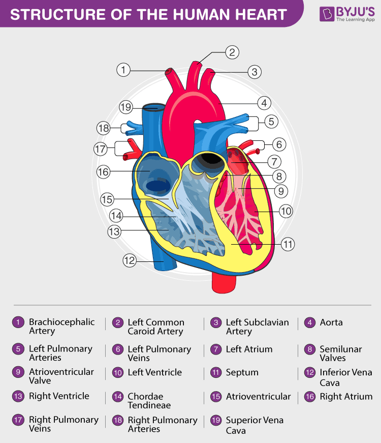
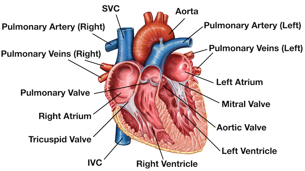
Draw And Label The Human Heart - It also has several margins: The four types of valves are: This will also help you to draw the structure and diagram of human heart. For this first step of our guide on how to draw a human heart, we will start with some outlines for the heart. Base (posterior), diaphragmatic (inferior), sternocostal (anterior), and left and right pulmonary surfaces. In this lecture, dr mike shows the two best ways to draw and label the heart! Drag and drop the text labels onto the boxes next to the diagram. Web cardiovascular heart diagram: Right, left, superior, and inferior: Web to draw the internal structure of the heart, start by sketching the 2 pulmonary veins to the lower left of the aorta and the bottom of the inferior vena cava slightly to the right of that. After reading this article you will learn about the structure of human heart. Web cardiovascular heart diagram: The upper two chambers of the heart are called auricles. What does the heart look like. Your heart sure does work hard, but that doesn’t mean you have to work hard to draw it! Find out about and describe the basic needs of animals, including humans, for survival (water, food and air) Selecting or hovering over a box will highlight each area in the diagram. 14 views 1 year ago. The upper two chambers of the heart are called auricles. Web this interactive atlas of human heart anatomy is based on medical illustrations and. Web diagram of the human heart (cropped).svg. A typical heart is approximately the size of your fist: The right margin is the small section of the right atrium that extends between the superior and inferior vena cava. Light pencil shading of the heart. Shading the upper sections of the heart in pen. This will also help you to draw the structure and diagram of human heart. Web to draw the internal structure of the heart, start by sketching the 2 pulmonary veins to the lower left of the aorta and the bottom of the inferior vena cava slightly to the right of that. Web explore the cardiovascular system of the human body with innerbody's interactive 3d anatomy models. Web what is the heart? Web the diagram of heart is beneficial for class 10 and 12 and is frequently asked in the examinations. In this lecture, dr mike shows the two best ways to draw and label the heart! In this interactive, you can label parts of the human heart. 41k views 1 year ago cardiovascular system. The heart features four types of valves which regulate the flow of blood through the heart. The user can show or hide the anatomical labels which provide a useful tool to create illustrations perfectly adapted for teaching. 244 × 240 pixels | 489 × 480 pixels | 782 × 768 pixels | 1,043 × 1,024 pixels | 2,086 × 2,048 pixels | 663 × 651 pixels. What does the heart look like. Original file (svg file, nominally 663 × 651 pixels, file size: Your heart contains four muscular sections ( chambers) that briefly hold blood before moving it. Selecting or hovering over a box will highlight each area in the diagram. 14 views 1 year ago.Heart Anatomy Labeled Diagram, Structures, Blood Flow, Function of

Heart Diagram with Labels and Detailed Explanation

Heart Anatomy Anatomy and Physiology
Base (Posterior), Diaphragmatic (Inferior), Sternocostal (Anterior), And Left And Right Pulmonary Surfaces.
Draw The Main Shape Of Your Human Heart Drawing.
Within The Triangle, Draw A Horizontal And Vertical Centerline To Split The Triangle Into Four Pieces.
12 Cm (5 In) In Length, 8 Cm (3.5 In) Wide, And 6 Cm (2.5 In) In Thickness.
Related Post:
