Cranial Nerves Drawing Face
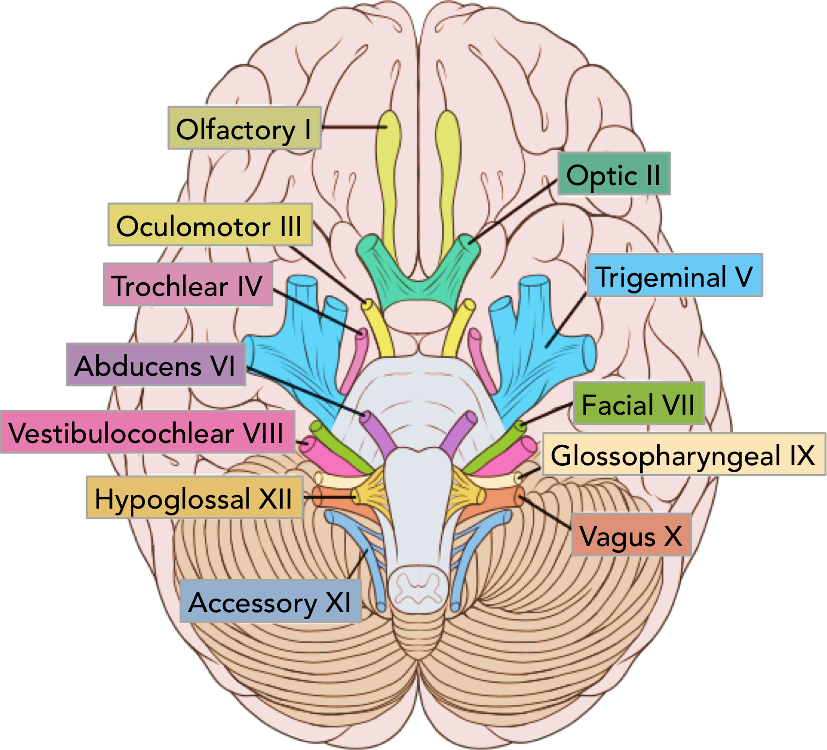
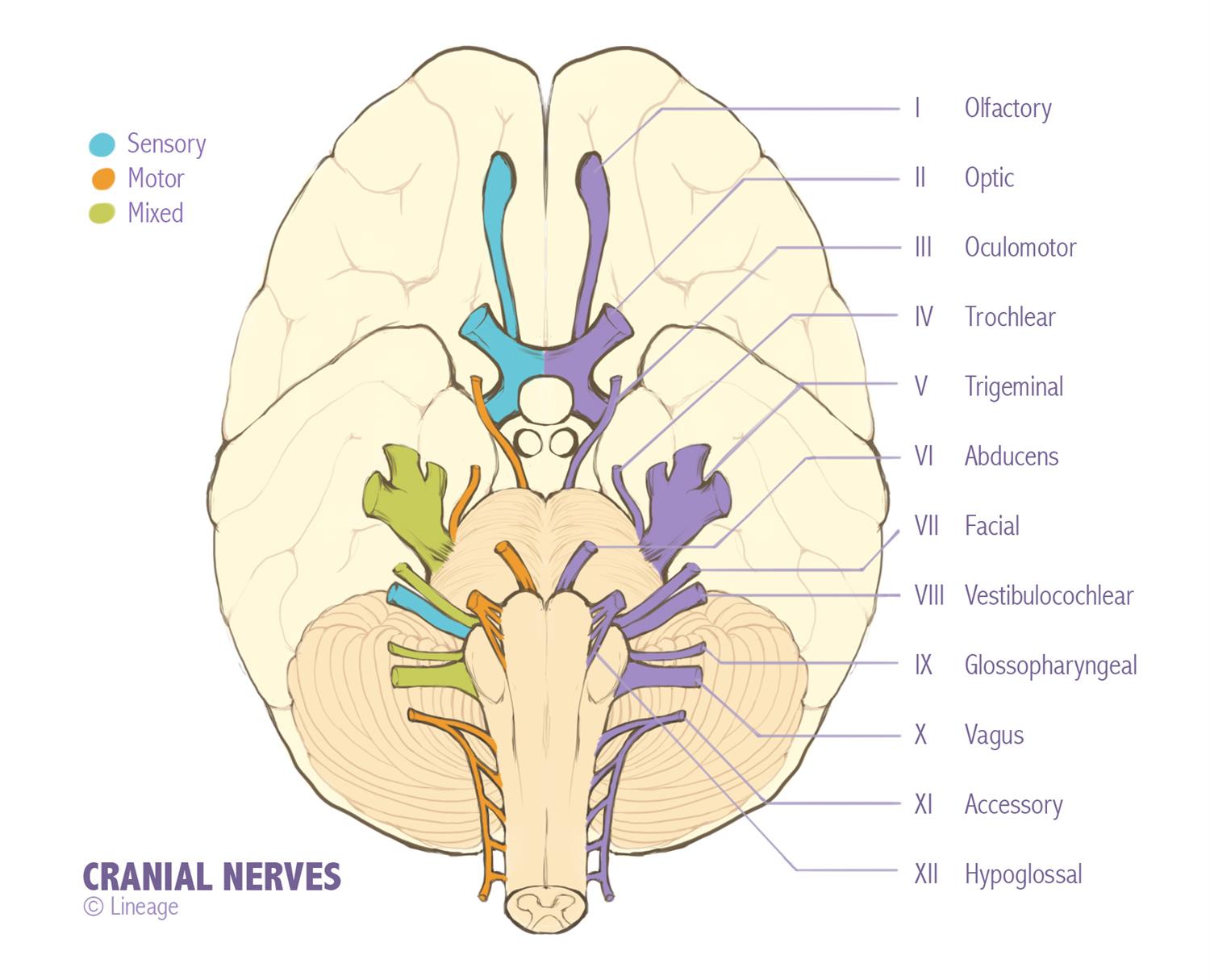
Cranial Nerves Drawing Face - Web easier way to remember the cranial nerves and their locations—by drawing a face and using numbers as the facial features. Web the facial nerve is the seventh cranial nerve (cn vii). Learn and reinforce your understanding of anatomy of the facial nerve (cn vii). A larger primary root carrying motor fibers and a smaller intermediate nerve carrying sensory and parasympathetic fibers. Both sides of the face should move the same way. Color each part according to the keys. Olfactory bulb and tract (purple) optic nerve and chiasma (dark green) oculomotor (dark blue) trochlear (gray) trigeminal (pink) abducens (orange) The human body has 12 pairs of cranial nerves that control motor and sensory functions of the head and neck. Web the facial nerve is associated with the derivatives of the second pharyngeal arch. Web the human body has 12 pairs of cranial nerves that control motor and sensory functions of the head and neck. Twelve pairs of nerves (the cranial nerves) lead directly from the brain to various parts of the head, neck, and trunk. The facial nerve is the seventh cranial nerve, and it is responsible for some taste as well as motor innervation to most muscles of the face. Travels through the base of your skull near the vestibulocochlear nerve, the eighth. Have him wrinkle his forehead, close his eyes, smile, pucker his lips, show his teeth, and puff out his cheeks. Accessory nerve (or spinal accessory nerve): Ability to taste and swallow. Web the human body has 12 pairs of cranial nerves that control motor and sensory functions of the head and neck. On the weakened side, the nasolabial fold is. Number the cranial nerves appropriately. Web the facial nerve is associated with the derivatives of the second pharyngeal arch. This article provides a pictorial overview of the imaging of cranial nerves, with a special focus on their anatomy and pathology. Web easier way to remember the cranial nerves and their locations—by drawing a face and using numbers as the facial. Web the 12 cranial nerves are pairs of nerves that start in different parts of your brain. The vestibulocochlear nerve, cn viii, transmits sound and balance information to the brain from the ear. Twelve pairs of nerves (the cranial nerves) lead directly from the brain to various parts of the head, neck, and trunk. You have two facial nerves, one on each side of your head. Web 2) an excel style chart outlining the nerve, fibers, innvervation, functions, and brainstem nucleus. Travels through the base of your skull near the vestibulocochlear nerve, the eighth cranial nerve, which helps you hear and maintain balance. This article provides a pictorial overview of the imaging of cranial nerves, with a special focus on their anatomy and pathology. Web the 7th (facial) cranial nerve is evaluated by checking for hemifacial weakness. Learn and reinforce your understanding of anatomy of the facial nerve (cn vii). On the weakened side, the nasolabial fold is depressed and the palpebral fissure is widened. Anatomy of the facial nerve (cn vii) videos, flashcards, high yield notes, & practice questions. Asymmetry of facial movements is often more obvious during spontaneous conversation, especially when the patient smiles or, if obtunded, grimaces at a noxious stimulus; Shoulder and neck muscle movement. Ability to move your tongue. In addition to motor fibers, this multitasking nerve also. It originates from the brainstem as two separate divisions;
Summary of the Cranial Nerves TeachMeAnatomy

Drawing Of The Face And Cranial Nerves Cranial+Nerves+On+Models+Labeled

Cranial Nerves Neurology Medbullets Step 1
Assess The Patient For Facial Symmetry.
30K Views 3 Years Ago #Cranialnerves #Mnemonic #Physiotutors.
This Video Is An Overview Of The Function Of The Following Cranial Nerves:1.
There Are Many Branches, Which Transmit A Combination Of Sensory, Motor And Parasympathetic Fibres.
Related Post: