Cranial Drawer Test Dog
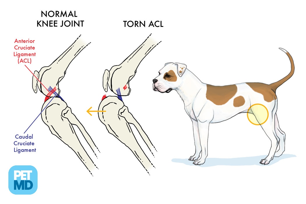
Cranial Drawer Test Dog - Web diagnosis of cranial cruciate ligament rupture is usually made by a positive cranial drawer sign. Sliding of the distal femur over the proximal tibia In general, radiographic images are used to visualize the instability of the stifle joint by tibial compression, to detect effusion and secondary osteoarthritic changes. Web welcome to our canine physiotherapy tutorial video, where we will guide you through two essential diagnostic tests for evaluating cranial cruciate ligament (. Some dogs are more relaxed in the standing position than when restrained in lateral recumbency. Web pain upon forced full extension of the stifle is a simple test that is suggestive of early crcld. (see figure 1 & cranial drawer & cranial tibial thrust tests.) Web specific palpation techniques that veterinarians use to assess the crcl include the ‘cranial drawer test’ and the ‘tibial compression test.’ these tests can confirm abnormal motion within the knee consistent with rupture of the crcl. Web definitive diagnosis of rupture of the ccl demands an assessment of stifle joint stability by means of the cranial “drawer” test, the tibial compression test, or both tests. Web a positive tibial compression test and cranial drawer test confirm cclr. Some dogs are more relaxed in the standing position than when restrained in lateral recumbency. (see figure 1 & cranial drawer & cranial tibial thrust tests.) Web the correct performance of either test is a learned skill, mastered only after much experience and practice on healthy dogs as well as those with partial or complete crclrs. Web a positive tibial. Web the correct performance of either test is a learned skill, mastered only after much experience and practice on healthy dogs as well as those with partial or complete crclrs. Web specific palpation techniques that veterinarians use to assess the crcl include the ‘cranial drawer test’ and the ‘tibial compression test.’ these tests can confirm abnormal motion within the knee. In a mature dog, a healthy, intact cranial cruciate ligament will not permit cranial tibial translation with the stifle held in extension or in flexion.3 in an immature dog, puppy laxity may permit a few millimeters of cranial and caudal tibial translation, but. Web the correct performance of either test is a learned skill, mastered only after much experience and. The cranial drawer test and tibial compression tests are important for assessing palpable instability. In a mature dog, a healthy, intact cranial cruciate ligament will not permit cranial tibial translation with the stifle held in extension or in flexion.3 in an immature dog, puppy laxity may permit a few millimeters of cranial and caudal tibial translation, but. Web the correct performance of either test is a learned skill, mastered only after much experience and practice on healthy dogs as well as those with partial or complete crclrs. Sliding of the distal femur over the proximal tibia In this test, the dog’s knee is slightly bent and anterior pressure is applied to the distal femur while posterior pressure is applied to the proximal tibia. Web a positive tibial compression test and cranial drawer test confirm cclr. The cranial drawer assessment is best done on the laterally recumbent animal. Some dogs are more relaxed in the standing position than when restrained in lateral recumbency. Web diagnosis of cranial cruciate ligament rupture is usually made by a positive cranial drawer sign. Web welcome to our canine physiotherapy tutorial video, where we will guide you through two essential diagnostic tests for evaluating cranial cruciate ligament (. Web specific palpation techniques that veterinarians use to assess the crcl include the ‘cranial drawer test’ and the ‘tibial compression test.’ these tests can confirm abnormal motion within the knee consistent with rupture of the crcl. Web specific palpation techniques that veterinarians use to confirm a problem with the ccl are the “cranial drawer test” and the “tibial thrust test.” these tests confirm abnormal motion in the knee and hence a rupture of the ccl. Web pain upon forced full extension of the stifle is a simple test that is suggestive of early crcld. Immature dogs are often misdiagnosed with crclr because they have greater than expected cranial drawer sign due to normal puppy laxity. Web definitive diagnosis of rupture of the ccl demands an assessment of stifle joint stability by means of the cranial “drawer” test, the tibial compression test, or both tests. In general, radiographic images are used to visualize the instability of the stifle joint by tibial compression, to detect effusion and secondary osteoarthritic changes.
Cruciate Disease The Cranial Drawer Test YouTube
Cranial Cruciate Ligament Medical Diagram Torn Knee Ligament in Dogs

Cranial Cruciate Ligament Disease in Dogs New England Veterinary Services
6 Evaluation Of Cd Signs And Ctt, Which Are Diagnostic Tests For Ccld, Should Be Performed During Examination.
Web To Test For Cranial Tibial Translation, Perform The Cranial Drawer Test (Figure 6).
The Examiner Stands Behind The Dog And Places A Thumb On The Caudal Aspect Of The Femoral.
(See Figure 1 & Cranial Drawer & Cranial Tibial Thrust Tests.)
Related Post: