Connective Tissue Drawing With Label
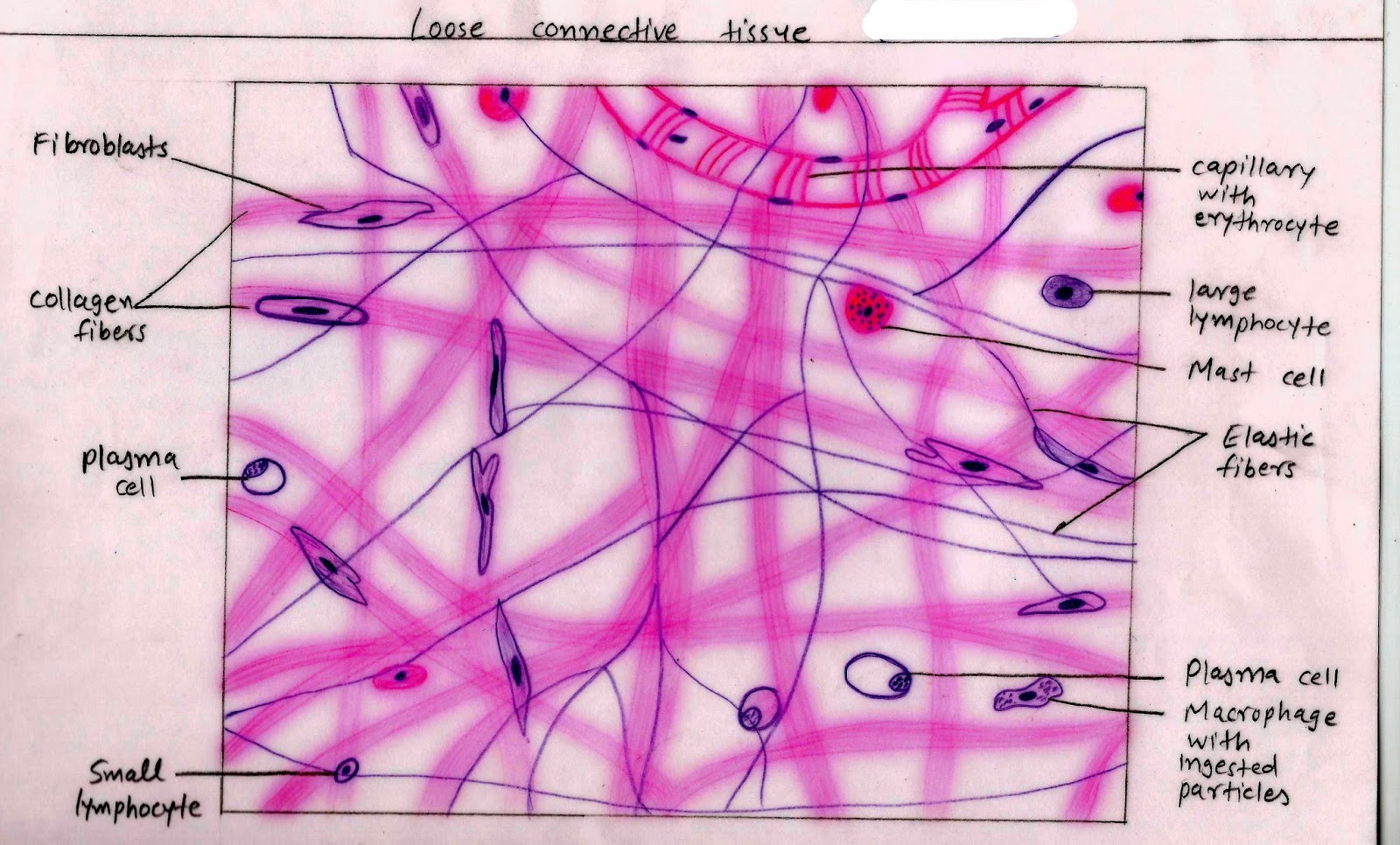
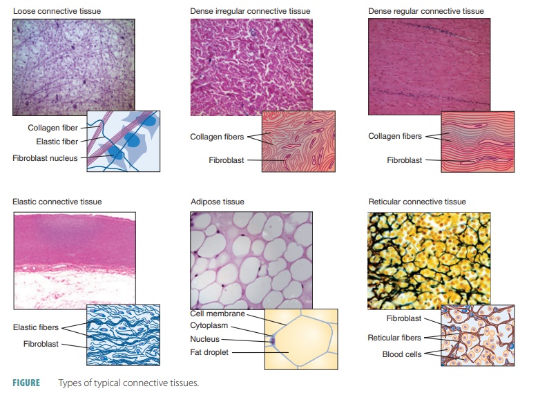
Connective Tissue Drawing With Label - Web in this micrograph of loose connective tissue of the tracheal mucosa numerous (labeled) cells of the connective tissue are present. Web our interactive anatomy tissue quizzes are the best way to make rapid progress, but these connective tissue quizzes with pictures are a great way to get started. It comprises a diverse group of cells that can be found in different parts of the body. Web in drawing images of connective tissue proper preparations seen under the microscope, it is important to simplify the visuals. O compare the molecular makeup, structural organization, location, and functions of the three main fiber types of connective tissue. This includes dense irregular connective tissue, cartilaginous tissue and bone tissue. As may be obvious from its name, one of the major functions of connective tissue. Connective tissue consists of three main components: List the major sarcomeric proteins involved with contraction. Describe the structure and function of skeletal muscle fibers. Indicate whether each figure represents a relaxed or. Web look no further than our connective tissue quizzes and diagram labeling exercises. Examples include adipose, cartilage, bone, blood, and lymph. Connective tissue proper includes loose connective tissue and dense connective tissue. Connective tissues also provide support and assist movement, store and transport energy molecules, protect against infections, and contribute to temperature. Cells, protein fibers, and an amorphous ground substance. Connective tissues also provide support and assist movement, store and transport energy molecules, protect against infections, and contribute to temperature homeostasis. List the major sarcomeric proteins involved with contraction. Describe the connective tissue layers surrounding skeletal muscle. Connective tissue proper and specialized connective tissue. Connective tissue is the tissue that connects or separates, and supports all the other types of tissues in the body. Web connective tissue is divided into four main categories: Connective tissues also provide support and assist movement, store and transport energy molecules, protect against infections, and contribute to temperature homeostasis. Web label the connective tissue in the figure. Note the. Bone, or osseous tissue, is a connective tissue that has a large amount of two different types of matrix material. Dense connective tissue is divided into 1) dense regular, 2) dense irregular, 3) elastic. Connective tissue is the tissue that connects or separates, and supports all the other types of tissues in the body. Connective tissues also provide support and assist movement, store and transport energy molecules, protect against infections, and contribute to temperature homeostasis. List the major sarcomeric proteins involved with contraction. Download pdf worksheet (blank) download pdf worksheet (labeled) Define a muscle fiber, myofibril, and sarcomere. Indicate whether each figure represents a relaxed or. The ecm is composed of a moderate amount of ground substance and two main types of protein fibers: O correlate morphology of resident and wandering ct cells with their locations and functions. Supporting connective tissue comprises bone and cartilage. We will examine those tissues in greater detail in lab 5 the appendicular skeleton & lab 6 the axial. Web in drawing images of connective tissue proper preparations seen under the microscope, it is important to simplify the visuals. Web some are solid and strong, while others are fluid and flexible. Both tissues have a variety of cell types and protein fibers suspended in a viscous. Many different cells contribute to the formation of connective tissues.
Connective Tissues Biology for Majors II
Dense Connective Tissue Structure
Connective Tissue Labeled
As May Be Obvious From Its Name, One Of The Major Functions Of Connective Tissue.
Web Connective Tissue Is A Term Used To Describe A Variety Of Types Of Tissues.
Web While The Various Connective Tissues Of The Body Are Diverse, They Share Numerous Structural And Functional Features That Explain Why They Are Subsumed Into A Single Tissue Category.
Loose Connective Tissue Is Divided Into 1) Areolar, 2) Adipose, 3) Reticular.
Related Post: