Chromatin Drawing
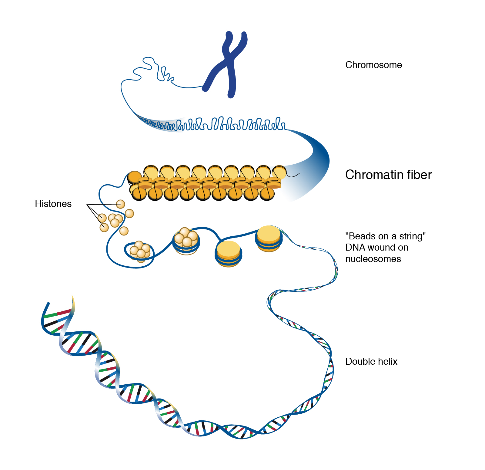
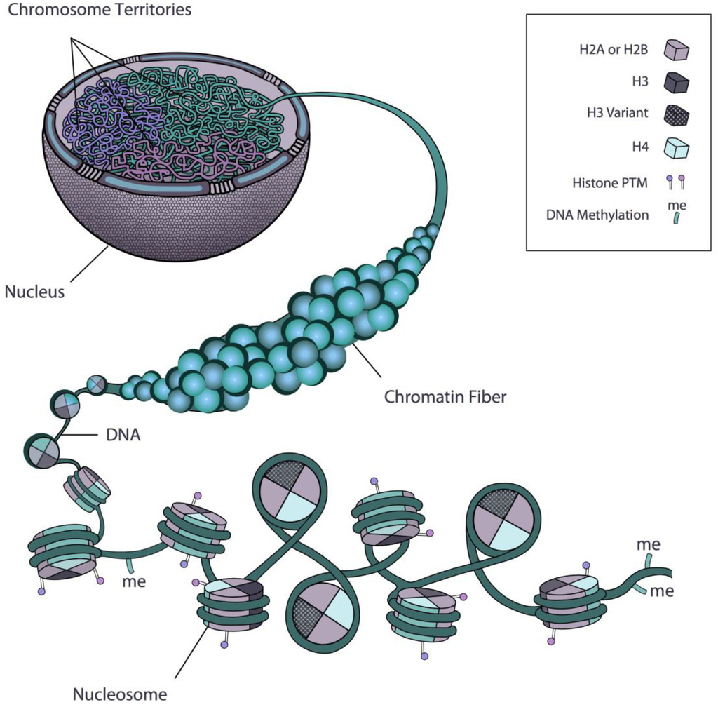
Chromatin Drawing - Tools to physically manipulate chromatin; Methods used to analysis chromatin. These 46 chromosomes are organized into 23 pairs: [1] the primary function is to package long dna molecules into more compact, denser structures. This review discusses the principles that drive the spatial architecture of chromatin, as well as genome‐wide‐binding patterns of chromatin proteins. Dna replication, transcription, and translation are key biological processes. Web mitosis consists of four basic phases: Web begin to move them towards opposite poles of the cell drawing. Prophase, metaphase, anaphase, and telophase. H1, h2a, h2b, h3 and h4. Web thus, within the nucleus, histones provide the energy (mainly in the form of electrostatic interactions) to fold dna. For most of the life of the cell, chromatin is decondensed, meaning that it exists in long, thin strings that look like squiggles under the microscope. Its prime function lies in the packaging of dna molecules in a very long denser. Phase, also called the first gap phase, the cell grows physically larger, copies organelles, and makes the molecular building blocks it will need in later steps. Transcription involves dna creating mrna, and translation converts mrna into proteins. Dna replication, transcription, and translation are key biological processes. Dna, the nucleosome, the 10 nm beads on a string chromatin fibre and the. Web mitosis consists of four basic phases: Web as dna repair, replication, and transcription involve active reorganization of chromatin, live visualization of chromatin motions will elucidate new functional mechanisms of these nuclear processes. For most of the life of the cell, chromatin is decondensed, meaning that it exists in long, thin strings that look like squiggles under the microscope. Early. In s phase, the cell synthesizes a complete copy of the dna in its nucleus. Early 20th century gene mapping even showed the relative location (locus) of genes on chromosomes. Cutting edge microscopy techniques to image chromatin organization with super resolution; In the s phase, the cell's dna is replicated. Chromatin is a mass of genetic material composed of dna and proteins that condense to form chromosomes during eukaryotic cell division. Computational imaging tools to interpret chromatin structure and dynamics; In this stage, the chromatin coils and condenses into chromosomes. Web various genome‐wide mapping techniques have begun to reveal that, despite the tremendous complexity, chromatin organization is governed by simple principles. [1] the primary function is to package long dna molecules into more compact, denser structures. Histones are small and positively charged proteins and are of 5 major types: Difference between chromosomes and chromatin. Prophase, metaphase, anaphase, and telophase. Different species have different numbers of chromosomes. For example, humans are diploid (2n) and have 46 chromosomes in their normal body cells. H1, h2a, h2b, h3 and h4. Histones are most abundant proteins in chromatin.
What are chromatin? Definition, Types and Importance biology AESL

Chromatin

Nucleosome Histone Chromatin
Web Chromatin Is Defined As A Complex Of Rna, Dna, And Protein Observed In Eukaryotic Cells.
The Major Structures In Dna Compaction:
Rather Each Chromosome Occupies A Spatially Limited, Roughly Elliptical Domain Which Is Known As A Chromosome Territory (Ct).
Web Thus, Within The Nucleus, Histones Provide The Energy (Mainly In The Form Of Electrostatic Interactions) To Fold Dna.
Related Post: