Cardiac Muscle Drawing
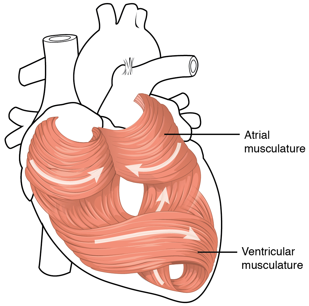
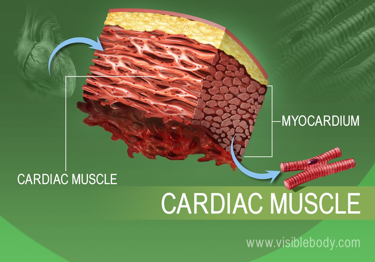
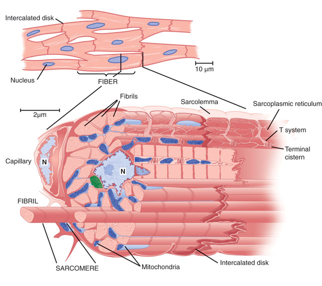
Cardiac Muscle Drawing - Cardiac muscle tissue, or myocardium, is a type of muscle tissue that forms the heart. Web introduction to the cardiac muscle tissue: David’s medical center demonstrated a new kind of heart. Cardiac muscle tissue contracts and releases. Web draw cardiac muscle tissue diagram easily with this video I will also enlist the functions and identification points of cardiac muscle. Cardiac muscle tissue is only found in your heart. Two cardiac muscle cell nuclei are indicated in the labelled image. The video describes the summary of the. The individual cardiac muscle cells are arranged in bundles that form a spiral pattern in the wall of the heart. It performs involuntary, coordinated contractions that allow your heart to pump blood. Cardiac muscle (or myocardium) makes up the thick middle layer of the heart. Identify and describe the components of the conducting system that distributes electrical impulses through the heart. 201 views 3 months ago easy science drawing. 1 waiting premieres may 2, 2023 #histology #anatomy #lpanatomy. Watch the video tutorial now. 1 waiting premieres may 2, 2023 #histology #anatomy #lpanatomy. Cardiac muscle is similar to skeletal muscle, another major muscle type, in that it possesses contractile units known as sarcomeres; 5.9k views 2 years ago #class 9 science : Cardiac muscle tissue is only found in your heart. 34k views 2 years ago #muscle #tissue #cardiac. The cardiac muscle or the myocardium forms the musculature of the heart. Watch the video tutorial now. These inner and outer layers of the heart, respectively, surround the cardiac muscle tissue and separate it from the blood. Its parts work together to move blood through your body in a coordinated way. Cardiac muscle cells ( cardiocytes or cardiac myocytes) make up the myocardium portion of the heart wall. Web keep exploring byju’s biology for more such exciting diagram topics. Many conditions can affect this organ and keep it from working well. The video describes the summary of the. Cardiac muscle, also known as heart muscle, is the layer of muscle tissue which lies between the endocardium and epicardium. The individual cardiac muscle cells are arranged in bundles that form a spiral pattern in the wall of the heart. These inner and outer layers of the heart, respectively, surround the cardiac muscle tissue and separate it from the blood. Web how to draw diagram of cardiac muscle step by step for beginners ! In the connective tissue between cardiac. A cardiac muscle cell typically has one nucleus located near the center. How to draw a muscle. After the end of the article, i will share the cardiac muscle histology drawing with you. By the end of this section, you will be able to: It is capable of strong, continuous, and rhythmic contractions that are automatically generated. Web cardiac muscle tissue is only found in the heart. Compare the effect of ion movement on membrane potential of cardiac conductive and contractile cells.
Musculo Corazon

Cardiac Muscle Structure

cardiac muscles properties morphology Medicine Hack
David’s Medical Center Demonstrated A New Kind Of Heart.
Watch The Video Tutorial Now.
Web How To Draw Cardiac Muscles Step By Step In A Very Easy Way || Type Of Muscles Tissue.
Web Cardiac Muscle Tissue, Also Known As Myocardium, Is A Structurally And Functionally Unique Subtype Of Muscle Tissue Located In The Heart, That Actually Has Characteristics From Both Skeletal And Muscle Tissues.
Related Post: