Brainstem Drawing
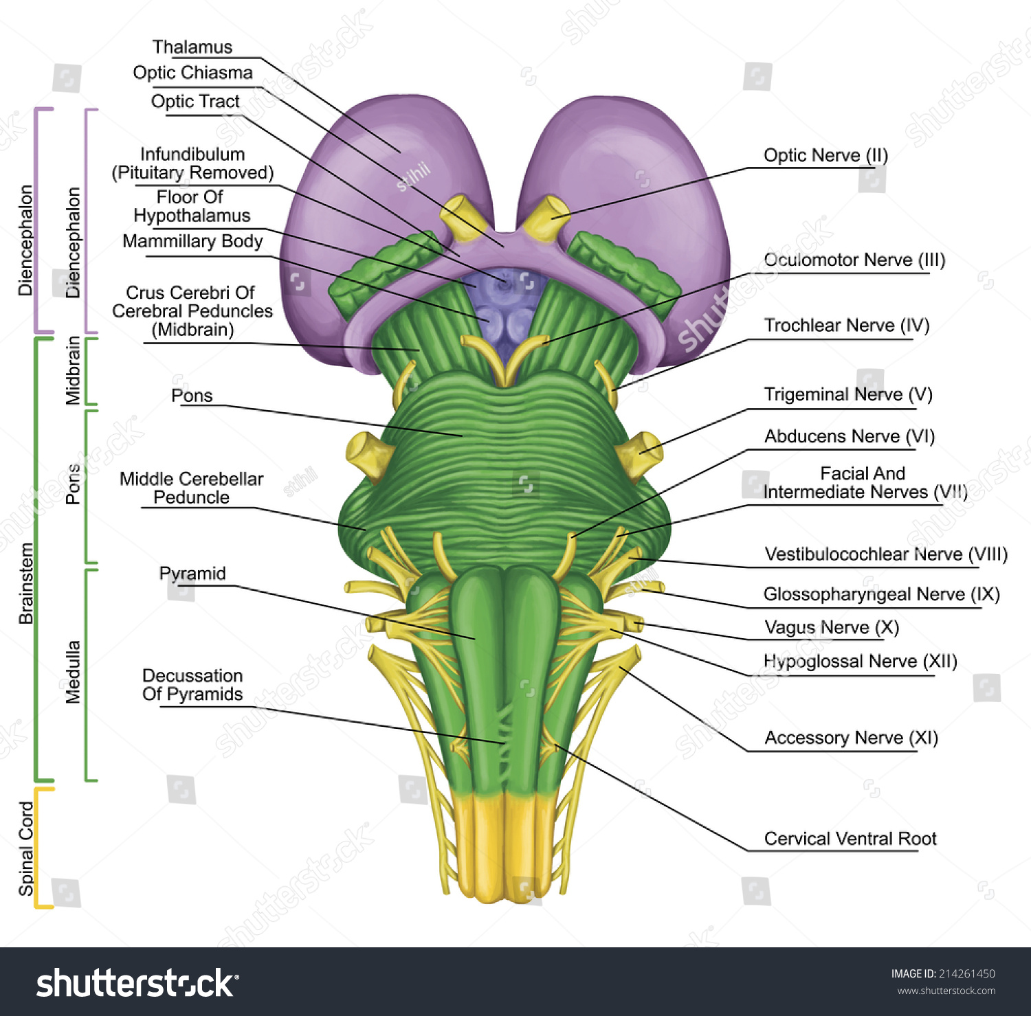
Brainstem Drawing - The brain stem consists of mesencephalon (midbrain), metencephalon, and myelencephalon. It is elegant, by explaining only that which can be detected by a basic neurological examination, and it is practical, by offering a system that can be easily remembered. The brainstem is the part of the brain that directly connects with the spinal cord. Watch the video tutorial now. Each of the three components has its own unique structure and function. Knowledge on ascending and descending nerve pathways, distribution of cranial nerve nuclei in. Brainstem [25:43] brainstem and related structures. Midsagittal drawing showing the main structures of the diencephalon. Web brainstem › drawing highlights. Web learn how to draw the brain stem and its anatomical features in this neuroanatomy tutorial video. Learn how to identify the main parts of the brain with labeling worksheets and quizzes. The medulla houses essential ascending and descending nerve tracts as well as brainstem nuclei. Draw a coronal view of the right brain. Web brainstem, area at the base of the brain that lies between the deep structures of the cerebral hemispheres and the cervical spinal. The brain stem consists of mesencephalon (midbrain), metencephalon, and myelencephalon. Web the medulla oblongata (medulla) is one of the three regions that make up the brainstem. The medulla houses essential ascending and descending nerve tracts as well as brainstem nuclei. We will discuss clinical cases from the midbrain, pons, and medulla and learn the anatomy of the brainstem around these. The medulla houses essential ascending and descending nerve tracts as well as brainstem nuclei. Web the medulla oblongata (medulla) is one of the three regions that make up the brainstem. Internal anatomy of the spinal cord in cross sections. Web in this drawing they're showing sensory information coming in through cranial nerves to the brainstem, to different nuclei in different. Here, we will learn a consolidated view of the major ascending (sensory) pathways from the cerebrum through the brainstem into the spinal cord. Internal anatomy of the spinal cord in cross sections. Let's start with an anterior view of the brainstem, which is how we commonly study the brainstem in anatomy lab. Supplementary motor nuclei (eg, the substantia nigra, pontine nuclei, and inferior olive). Knowledge on ascending and descending nerve pathways, distribution of cranial nerve nuclei in. 7.2k views 10 years ago. Learn how to identify the main parts of the brain with labeling worksheets and quizzes. Web drawing on experimental data, mathematical modeling and theory, the scientists make the case that bursts of beta rhythms control cognition in the brain by regulating where and when higher gamma. Web in this episode, we will try to learn the brainstem anatomy from a clinical angle. These cranial nerves are mostly performing these functions in the head and the neck. We will discuss clinical cases from the midbrain, pons, and medulla and learn the anatomy of the brainstem around these cases. Web the medulla oblongata (medulla) is one of the three regions that make up the brainstem. Draw a coronal view of the right brain. Cranial nerve nuclei, part 1. Learn the general anatomy of the brainstem by using a compressed composite of all three of its axial levels: Anatomy of the brain (sagittal view) the cerebellum and brainstem are a testament to the fact that good things do come in small packages, so this article is an overview of their anatomy.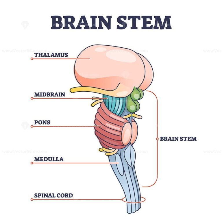
Brain stem parts anatomical model in educational labeled outline
/GettyImages-1092334754-fd0644493b3148288970e38fd26aead0.jpg)
Brainstem Anatomy, Function, and Treatment
Brainstem Ventral View Posterior Part Brain Stock Illustration
Web The Present Work Provides A Topographic Atlas Of The Human Brainstem Composed Of 45 Anatomical Plates, Each Containing A Pair Of Adjacent Sections Stained With Cresyl Violet And Luxol Fast Blue To Help Delineating Brainstem Nuclei.
Each Of The Three Components Has Its Own Unique Structure And Function.
Web The Agency Changed Course Earlier This Month After Shupe Appealed, Granting Her Copyright Registration For Ai Machinations:
Web The Brainstem, Positioned At The Base Of The Posterior Region Of The Brain, Functions As The Critical Point Between The Central/ Peripheral Nervous Systems.
Related Post: