Bile Duct Drawing
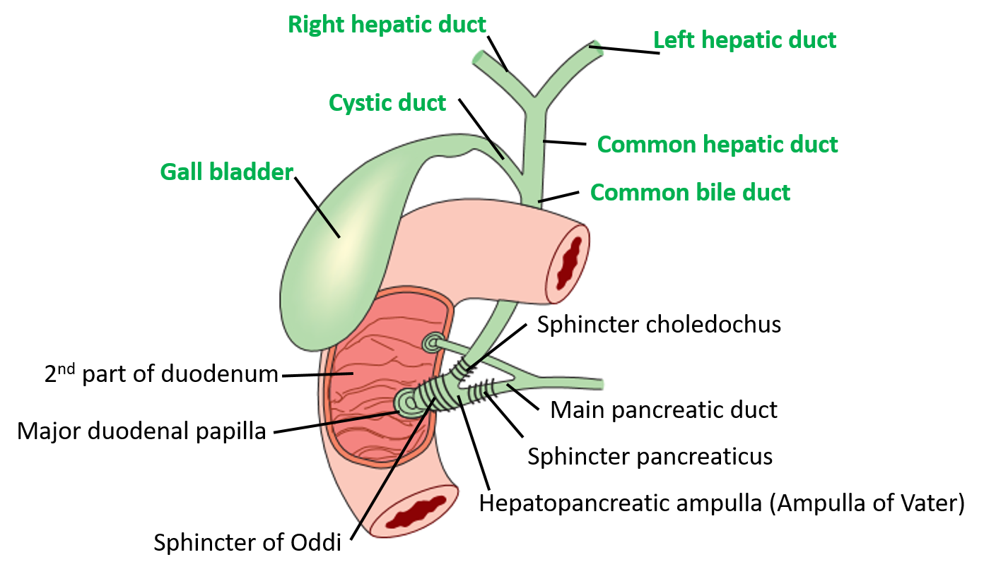
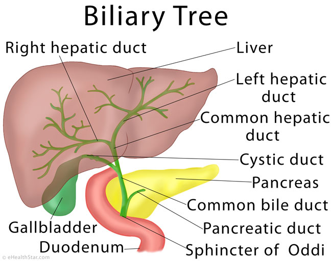
Bile Duct Drawing - Neurovascular supply and lymphatic drainage of the gallbladder. Web anatomy of the intrahepatic bile ducts; This is a small, hollow tube that functions to transport bile. Web a bile duct drain is a procedure that involves opening up obstructions or treating holes in the biliary system. Left and right hepatic ducts, 4. What is a bile duct drain? Web the biliary tree is a complex network of conduits that begins with the canals of hering and progressively merges into a system of interlobular, septal, and major ducts which then coalesce to form the extrahepatic bile ducts, which finally deliver bile. A bile duct obstruction is a blockage in your bile ducts. A normal gallbladder will have a thin, barely perceptible wall, and will not appear dilated. Drawing of the biliary system with the liver, biliary tree (bile ducts), common bile duct, gallbladder, pancreas, duodenal papilla, main pancreatic duct, and duodenum labeled. Drawing of the biliary system, with the liver, gallbladder, duodenum, pancreatic duct, common bile duct, pancreas, cystic duct, and hepatic ducts. When the liver cells secrete bile, it is collected by a system of ducts that flow from the liver through the right and left hepatic ducts. A bile duct drain, also called biliary drainage, can. In hepatic surgery, a. Some patients have calcified gallstones that are visible on ct and look like small bright dots. Drawing shows the extrahepatic bile ducts, including the common hepatic duct (perihilar region) and the common bile duct (distal region). Anatomy of the extrahepatic bile ducts; What is a bile (biliary) duct obstruction? This explains why secretin does not increase mannitol clearance. Web 135 kb | 600 x 951. 742 kb | 2025 x 2100. Web the common bile duct is formed by the junction of the cystic and hepatic ducts; Drawing of the biliary system with the liver, biliary tree (bile ducts), common bile duct, gallbladder, pancreas, duodenal papilla, main pancreatic duct, and duodenum labeled. There is significant variation in the. Inset of an enlarged biliary system with the. Some patients have calcified gallstones that are visible on ct and look like small bright dots. Web 135 kb | 600 x 951. It then runs in a groove near the. Web anatomy of the intrahepatic bile ducts; Bile, required for the digestion of food, is secreted by the liver into passages that carry bile toward the hepatic duct, which joins with the cystic duct (carrying bile to and from the gallbladder) to form the common bile duct, which opens into the intestine. The organs and bile ducts together form your biliary system. Gallstones are the most common cause of bile duct obstructions. Also shown is the common hepatic duct, gallbladder, cystic duct, common bile duct, pancreas, ampulla of. Web the transportation of bile follows this sequence: What is a bile (biliary) duct obstruction? Left and right hepatic ducts, 4. This is a small, hollow tube that functions to transport bile. Bile ducts are tiny canals that connect some of the organs in your digestive system. Drawing of the biliary system with the liver, gallbladder, pancreas, duodenum, bile ducts, cystic duct, common bile duct, and pancreatic duct labeled. Bile canaliculi unite to form segmental bile ducts which drain each liver segment.
Extrahepatic Biliary Apparatus Anatomy QA

Liver Anatomy, Location and Function eHealthStar

Anatomy of the Biliary System with Labels Media Asset NIDDK
Web Figure 21.7.4 Is A Drawing Showing The Anterior View Of The Gallbladder, Part Of The Liver Superior To The Gallbladder, And The Duct Connecting The Liver To Gallbladder, Eventually Becoming The Common Bile Duct.
This Explains Why Secretin Does Not Increase Mannitol Clearance.
The Biliary System Is Comprised Of A System Of These Ducts, Which Flow From The Liver To The Gallbladder For Storage And Then Into The Small Intestine (Duodenum).
These Ducts Ultimately Drain Into The Common Hepatic Duct.
Related Post: